Biology · Skim version
The Cell Theory and Its Evidence
Read the full lesson → Skim the whole course →
The guiding idea
Robert Hooke saw tiny boxes in a slice of cork in 1665, so why did it take about 175 more years to claim that plants and animals alike are made of cells?
The cell theory, that every living thing is made of cells and every new cell comes from a cell, needed more than better tools: a tool shows a detail only if the detail is larger than the tool's limit, and the theory also took comparing many living things and testing where new cells come from.
From the film: What a Microscope Can Show
- Magnification makes an image bigger; resolution decides how much detail it holds.
- An ordinary light microscope cannot separate two points closer than about 0.2 micrometers, however much you enlarge the image.
- A tool shows a detail only if the detail is larger than the tool's limit, so a whole cell can be clear while its parts are not.
- Hooke's cork showed the walls of dead cells from one plant, which was not enough to claim that all life is made of cells.
From the film: Where Do New Cells Come From?
- Schleiden and Schwann compared many plant and animal tissues and found the same cellular plan.
- They also thought new cells could form inside a structureless fluid, like a crystal, with no cell dividing, and that part was wrong.
- Remak published evidence in 1852 that new cells form when existing cells divide, and Virchow spread the idea in 1855: every cell comes from a cell. That rule is about cells living today; how the very first cells arose is a separate question.
- The cell theory kept its core and replaced the wrong part with a corrected idea, which is how a well-tested scientific theory changes.
-
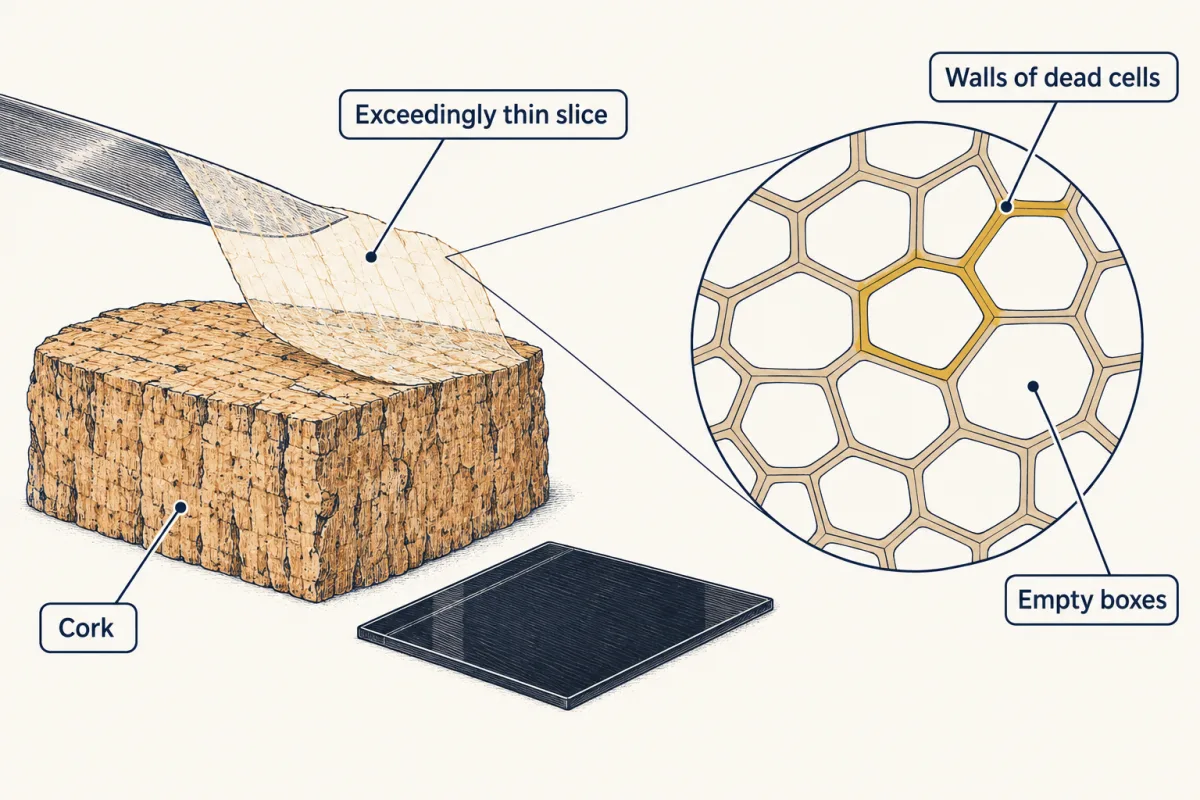
Hooke's thin slice of cork showed rows of tiny walled boxes, the walls of dead cells. AI-generated illustration -
Explore: zoom in and see what appears
-
Explain: seeing smaller things
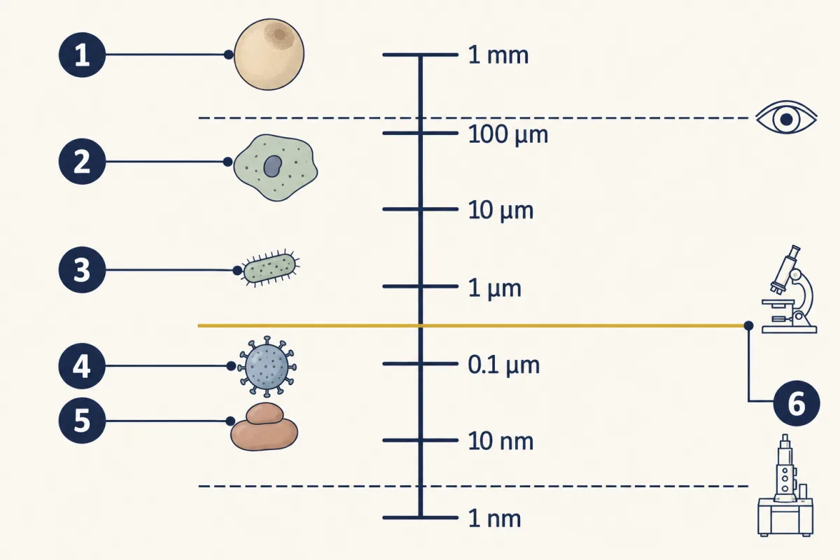
- frog egg
- cheek cell
- bacterium
- virus
- ribosome
- limit of the light microscope
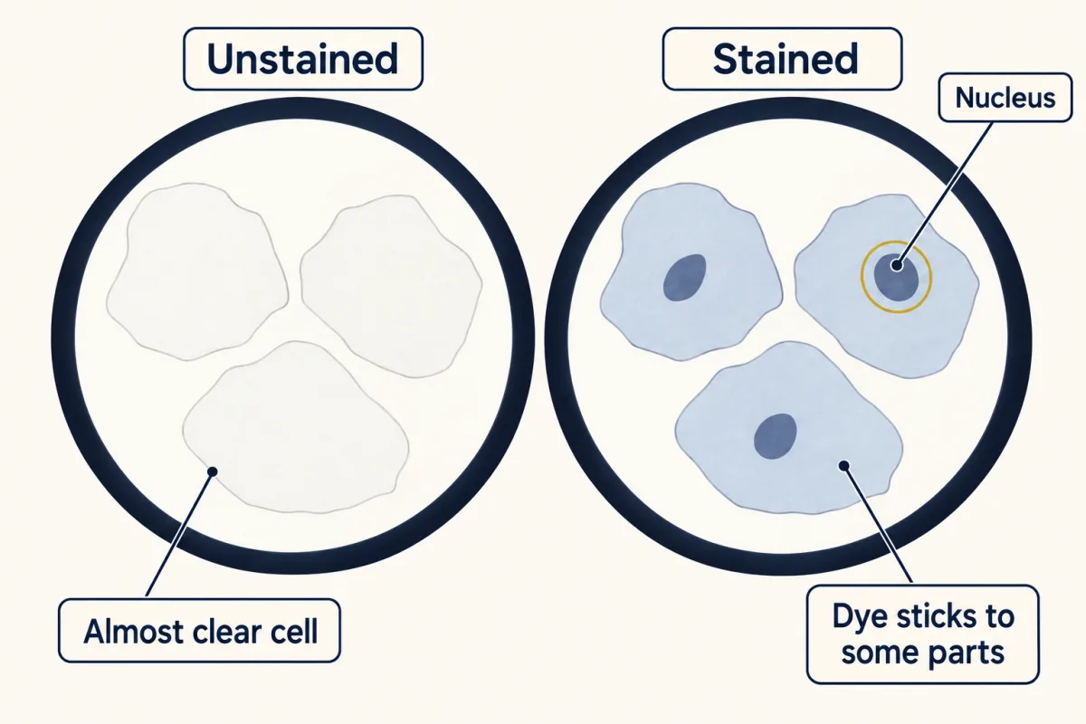
The same cells unstained and stained: the stain makes the nucleus stand out. AI-generated illustration -
Explain: the six ideas of the cell theory
-
Explain: a theory that grew and changed
-
Elaborate: tools and discoveries over time
-
Evaluate: check your thinking
-
Going deeper
Where this lesson fits
Before thisReview first
- What Am I Made Of?This course · Lesson 2
The sixth idea says cells share the same main molecules, which this lesson names and explains.
After thisStudy next
- The Cell Cycle and MitosisThis course · Lesson 10
It shows how an existing cell divides into two cells, the process behind the idea that all cells come from cells.
- Living or Not? Viruses and BacteriaThis course · Lesson 12
It tests the cell theory on viruses, which are not made of cells.
- Organelles as a SystemThis course · Lesson 7
It uses the finer cell parts that electron microscopes later revealed.
Elsewhere on the siteFilms, simulations and method films in other courses
- Zoom into a dropSimulation · Life Science
Zooming into pond water shows which things sit below the eye's size limit and need a microscope.
- Electron microscopyMethod film
It shows how electron beams reveal cell parts that light microscopes blur.
Suggested by an AI model from the lessons themselves, then checked. How we use AI