Cell Signaling · Skim version
Wnt: β-Catenin and Planar Polarity
Read the full lesson → Skim the whole course →
The guiding idea
Why would a cell make a protein all day only to destroy it?
Why does a cell make β-catenin all the time and destroy it all the time?
-
The name: a mouse tumour gene and a fly gene were one gene
-
The idea: β-catenin is made all the time and destroyed all the time
-
What Wnt changes: it stops the destruction
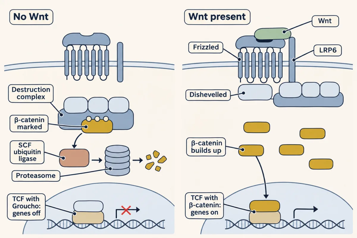
Without Wnt, the destruction complex marks β-catenin, SCF tags it with ubiquitin and the proteasome destroys it, while TCF holds Groucho and keeps target genes off. Wnt brings Frizzled and LRP6 together and pulls the complex to the receptor, so new β-catenin survives, builds up and joins TCF in the nucleus to switch genes on. How the complex is stopped is still argued. AI-generated illustration -
Wnt in disease: a pathway stuck on
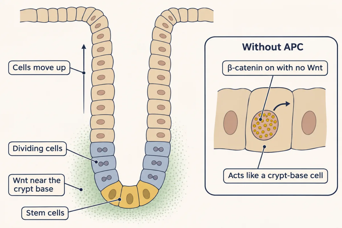
Stem cells at the base of the gut's crypts, and the dividing cells above them, depend on Wnt. A cell that loses APC keeps β-catenin and TCF switching genes on with no Wnt, so it behaves as if it had never left the crypt base. AI-generated illustration -
The other Wnt pathway: planar cell polarity tells a cell which side is which
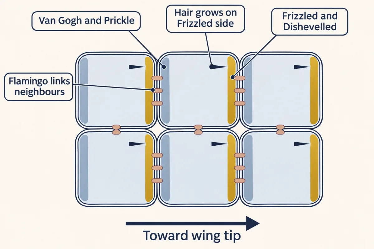
In each fly wing cell, Frizzled and Dishevelled gather on the edge toward the wing tip and Van Gogh and Prickle on the opposite edge, with Flamingo linking each cell to the next. The hair grows from the Frizzled side, so every hair points the same way, and none of this uses β-catenin or TCF. AI-generated illustration -
Going deeper
Where this lesson fits
Before thisReview first
- Ubiquitin and Regulated Proteolysis as SignalsThis course · Lesson 8
It gives the general rule that a ubiquitin ligase marks a protein for the proteasome, which the destruction complex uses on β-catenin.
- Adaptors, Docking Proteins and ScaffoldsThis course · Lesson 10
Axin works as a scaffold holding kinases next to their substrate, an idea that lesson teaches.
- PI 3-Kinase, Akt and InsulinThis course · Lesson 20
You need GSK3 as the kinase Akt switches off to see why insulin does not raise β-catenin.
After thisStudy next
- Hedgehog and the Primary CiliumThis course · Lesson 26
It follows another secreted, lipid-modified signal that uses a Frizzled-like receptor to steer embryo pattern.
- Oncogenes: When the Switch SticksThis course · Lesson 41
It takes the stuck-on switch idea further, with mutations that leave a pathway active without its signal.
- The Shape of a Signal: Size, Duration, Frequency and PlaceThis course · Lesson 35
It asks how cells read signal duration and place, including how one pathway gives different outputs.
Elsewhere on the siteFilms, simulations and method films in other courses
- Binding assays: immunoprecipitation, pull-down and gel shiftMethod film
Showing that Axin, APC and β-catenin sit in one complex relies on pulling one partner down with another.
- Gel electrophoresis, SDS-PAGE and western blotMethod film
Reading β-catenin level and its phosphorylated form in cell extracts uses this blot.
Suggested by an AI model from the lessons themselves, then checked. How we use AI