Physiology · Skim version
Ventilation, Perfusion and Gas Exchange
Read the full lesson → Skim the whole course →
The guiding idea
A resting adult moves about 6 litres of air and 5 litres of blood through the lungs every minute, yet neither number tells you what the blood leaves with. What does is whether air and blood arrive at the same places in the same proportion, and when they do not, gas exchange suffers.
What leaves the lungs in the blood depends on how well air and blood are matched, unit by unit, in a ratio of ventilation to perfusion.
-
One ratio sets each unit’s gas
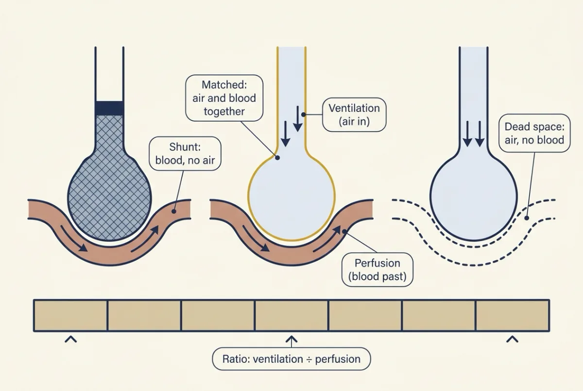
Every lung unit sits somewhere on one line, set by its ventilation divided by its perfusion: a shunt gets blood but no air, dead space gets air but no blood, and real units fall in between. AI-generated illustration -
Partial pressure is not content
-
Two units: why a spread costs oxygen
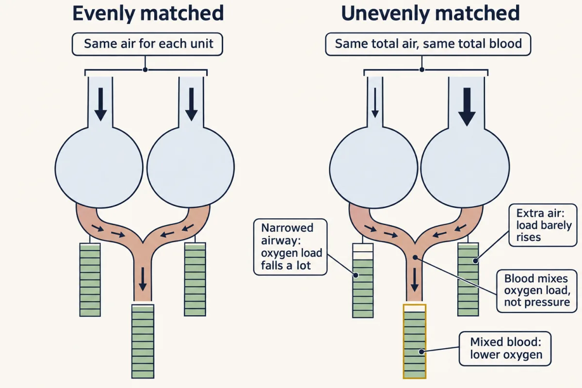
With the same total air and blood, uneven matching lowers arterial oxygen: the over-ventilated unit can add almost nothing because hemoglobin is already nearly full, while the under-ventilated unit loses a lot, and blood mixes the oxygen it carries, not its pressure. AI-generated illustration -
Measuring the gap: alveolar PO2 and the A-a difference
-
Dead space: the high end, measured
-
Hypoxic lungs constrict their own arteries
-
Top and bottom of the upright lung
-
Sorting low arterial oxygen with three numbers
-
Going deeper
Where this lesson fits
Before thisReview first
- Lung Mechanics and SurfactantThis course · Lesson 12
It gives the pressures that move air, which this lesson assumes when it splits ventilation among lung units.
- Smooth Muscle and the Myogenic ResponseThis course · Lesson 4
Hypoxic constriction of pulmonary arteries is built on smooth muscle contraction and its calcium and potassium-channel steps.
- Resistance, Compliance and Local Blood FlowThis course · Lesson 8
Hypoxia dilates systemic vessels, and you need that contrast and the resistance idea to see why lung vessels constrict.
After thisStudy next
- Control of Breathing, Altitude and DivingThis course · Lesson 14
It uses arterial PCO2 and alveolar oxygen to show how breathing is driven, and how altitude changes it.
- Exercise: The Whole Body at WorkThis course · Lesson 30
It asks which link in oxygen delivery limits uptake, using the matching and diffusion ideas from here.
- Pregnancy, Birth and LactationThis course · Lesson 29
The fall in lung vascular resistance at birth uses release of hypoxic constriction as the lungs fill with air.
Elsewhere on the siteFilms, simulations and method films in other courses
- The Last Gas the Blood TouchesFilm · Organismal Biology
It shows oxygen and carbon dioxide crossing exchange surfaces by partial pressure difference, the step this lesson builds on.
- Area, thickness and rateSimulation · Organismal Biology
It lets you vary area, thickness and rate to see how exchange depends on the partial pressure difference.
- Why Blood Needs an S-Shaped CarrierFilm · Organismal Biology
It shows how dissolved oxygen differs from hemoglobin-bound oxygen, the partial pressure versus content point used here.
Suggested by an AI model from the lessons themselves, then checked. How we use AI