Cell Signaling · Skim version
Sensing the World: Vision, Smell, Nitric Oxide and cGMP
Read the full lesson → Skim the whole course →
The guiding idea
Your rods report light by turning a current down. Why would a sense run backwards?
How does a rod cell turn one photon into a nerve signal, and why does it do so by destroying a messenger that was already there?
-
The idea: one bond turns, and everything after it is dark
-
A cascade that runs backwards: light makes cGMP fall
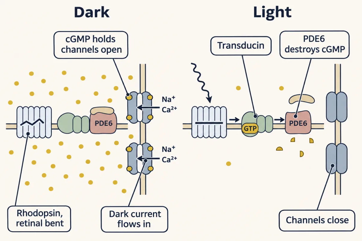
In the dark, cGMP holds the rod's channels open and a steady current flows in. Light straightens retinal, active rhodopsin switches on transducin, transducin frees PDE6, PDE6 destroys cGMP, and the channels close: the signal is a messenger that falls. AI-generated illustration -
Recovery: four things have to be undone
-
Color and smell: the same plan with more receptors
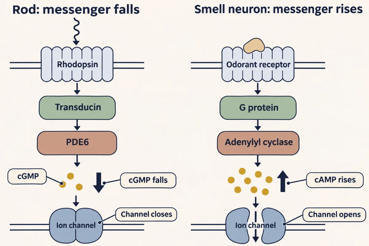
The eye and the nose use the same plan in opposite directions. In the rod, a G protein switches on PDE6, which breaks cGMP down, so cGMP falls and channels close; in a smell neuron, a G protein switches on adenylyl cyclase, cAMP rises and a related channel opens. AI-generated illustration -
A gas that relaxes arteries: nitric oxide raises cGMP
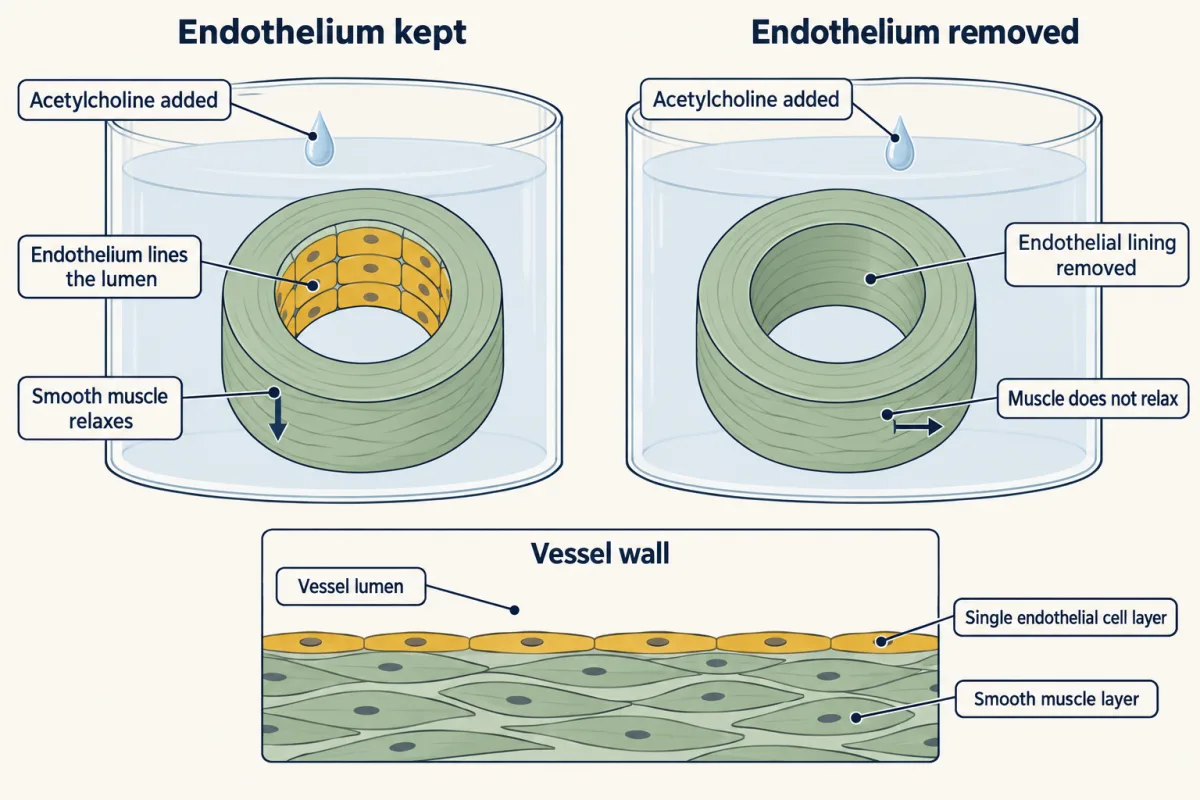
Furchgott and Zawadzki mounted rings of rabbit aorta in an organ bath. Acetylcholine relaxed rings that kept their endothelium and failed to relax rings where it had been rubbed off, so the lining was sending something to the muscle beneath it. AI-generated illustration
Where this lesson fits
Before thisReview first
- GPCRs and Trimeric G ProteinsThis course · Lesson 12
Rhodopsin and olfactory receptors are GPCRs that switch on G proteins, which you need before following either cascade.
- GTPase Switches: The TimerThis course · Lesson 7
Transducin's GTP-for-GDP exchange and its timed hydrolysis are the GTPase timer used to start and stop the rod cascade.
- Turning the Signal Off: Arrestins, RGS Proteins and PhosphodiesterasesThis course · Lesson 16
Rhodopsin kinase, arrestin, RGS proteins and phosphodiesterases are the shut-off parts the rod recovery relies on.
After thisStudy next
- Ion Channels as ReceptorsThis course · Lesson 30
It takes further the cyclic-nucleotide-gated channels and how channels turn signals into electrical responses.
- The Shape of a Signal: Size, Duration, Frequency and PlaceThis course · Lesson 35
It builds on the calcium feedback and adaptation seen in the rod to show how cells read signal size and duration.
Elsewhere on the siteFilms, simulations and method films in other courses
- Centre and surroundSimulation · Neuroscience
The retina lesson's simulation shows how a rod hyperpolarizes in light and releases less glutamate, the result of this cascade.
- One Cell CountsFilm · Neuroscience
It shows photon-driven closing of cGMP-gated channels and the falling glutamate release that follows.
- A Current That Light Can Use UpFilm · Sensory Systems
It shows how calcium feedback sets photoreceptor gain, the adaptation this lesson explains.
- One in Two ThousandFilm · Sensory Systems
It shows the one-receptor-per-neuron choice among a large gene family that this lesson compares with vision.
Suggested by an AI model from the lessons themselves, then checked. How we use AI