Biochemistry · Skim version
Protein Three-Dimensional Structure: The Sequence Sets the Shape
Read the full lesson → Skim the whole course →
The guiding idea
One sequence, one working shape. Change a single letter and the shape, and the job, can break.
A protein's amino acid sequence, in a given environment, sets its three-dimensional shape, and that shape sets its job.
-
One sequence, one shape
-
The proof: Anfinsen’s refolding experiment
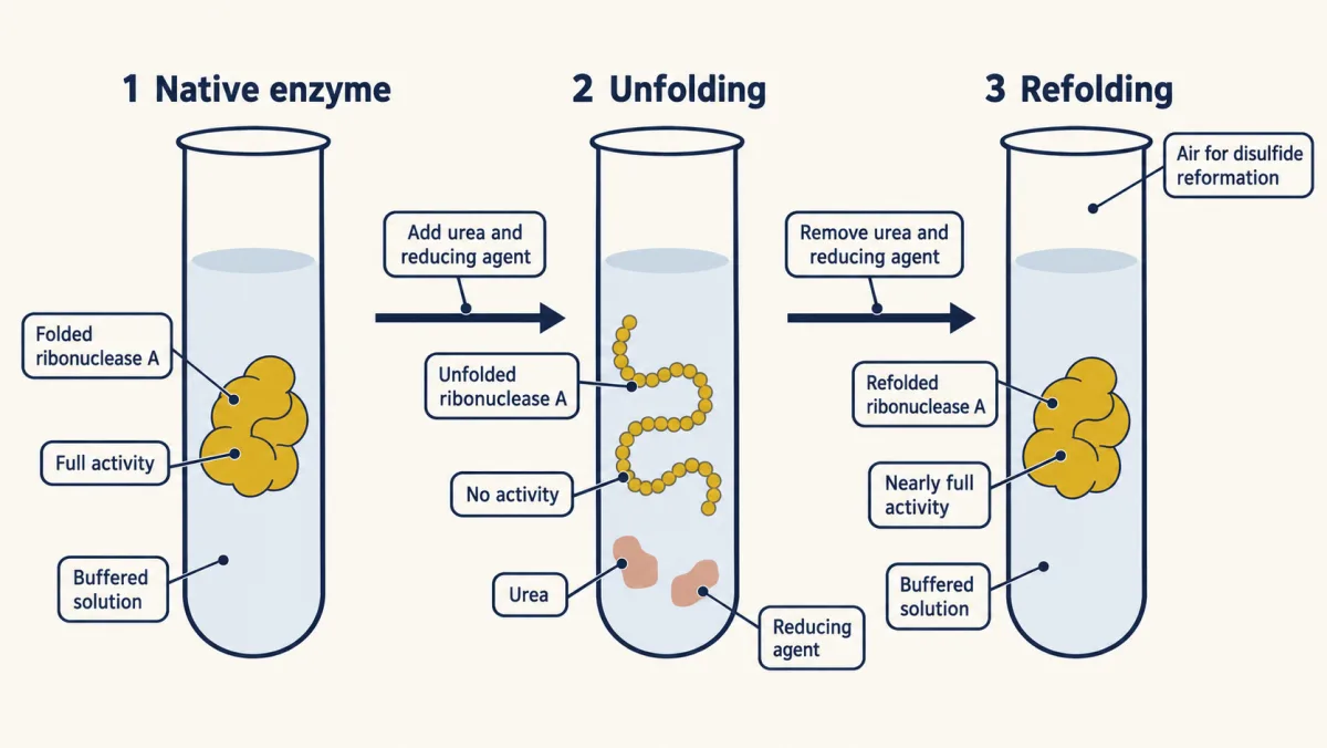
Anfinsen unfolded ribonuclease with urea and a reducing agent, so it lost all activity, then washed the chemicals away and the enzyme refolded on its own to full activity: the sequence alone holds the fold. AI-generated illustration -
The flat peptide bond: two hinges per residue
-
The map of allowed shapes: the Ramachandran plot
-
Helices and sheets: the backbone bonds to itself
-
The hydrophobic core: water pushes oil inward
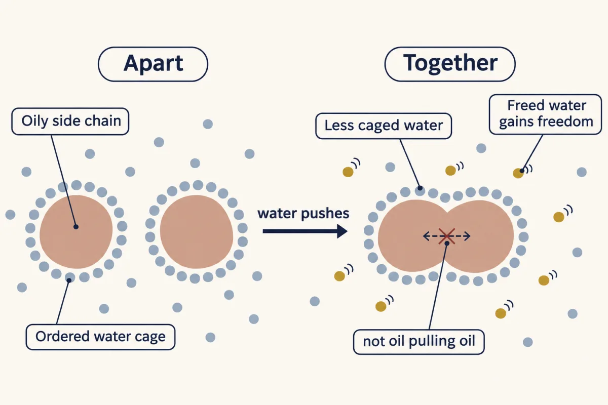
The hydrophobic effect. Water around an oily side chain is held in an ordered cage. Bring two oily groups together and less surface faces the water, so caged water is set free. That gain in the water's freedom drives the oily groups together: they are pushed together by water, not pulled together by each other. AI-generated illustration -
Between chains: quaternary structure
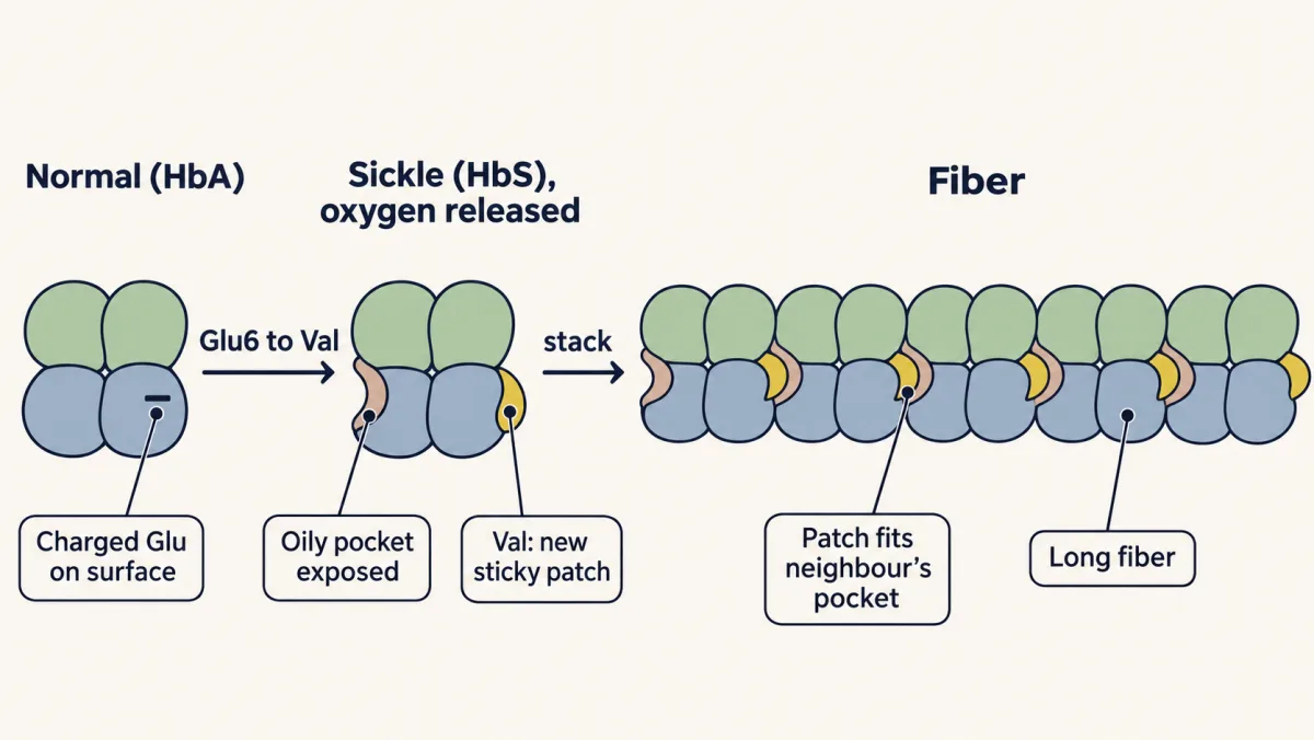
Sickle cell is the hydrophobic rule working between molecules. Valine replaces a charged glutamate on the surface of beta-globin, making a sticky patch. Once oxygen is released, an oily pocket is exposed, and the patch on one hemoglobin docks into the pocket of the next, stacking the molecules into long fibers. AI-generated illustration -
Only just stable: the melt
-
Finding the fold fast: Levinthal’s paradox and the funnel
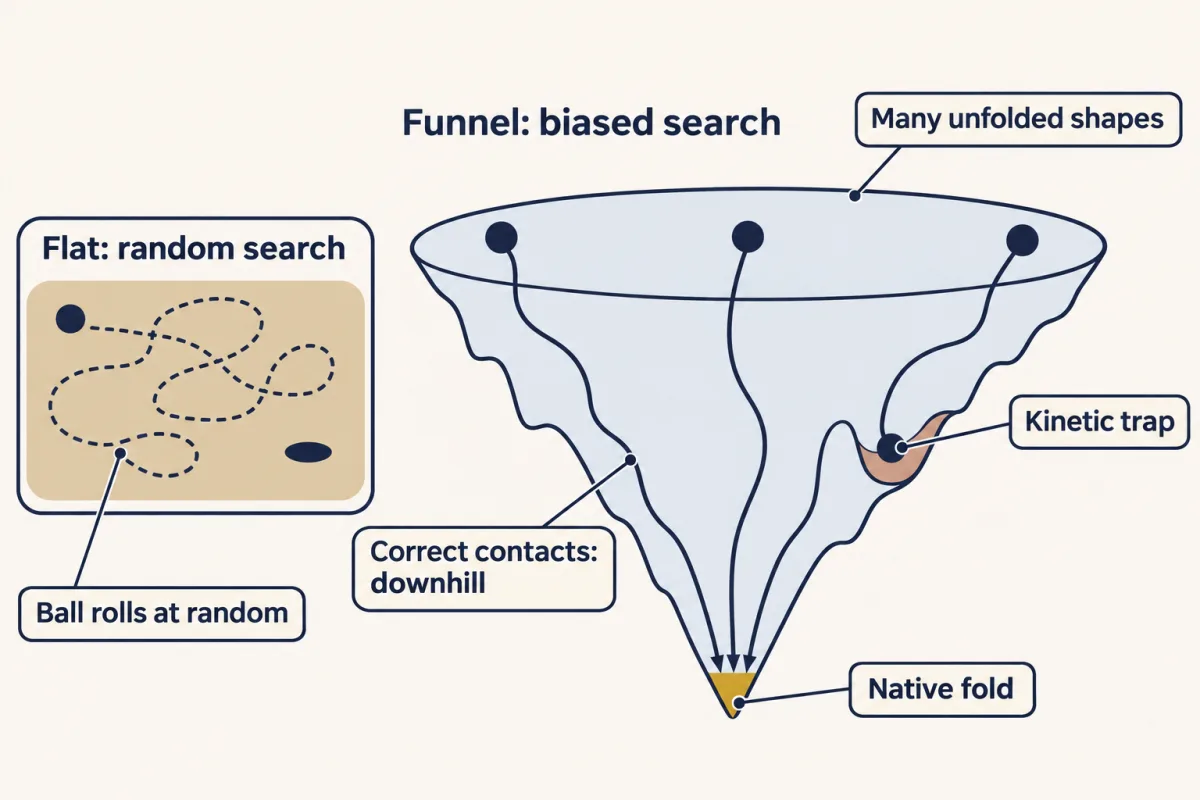
Why folding is fast. On a flat landscape a chain would search at random and almost never find its fold. Real chains fold on a funnel: once the first contacts are in place, native-like contacts lower the energy a little at each step, so wherever a chain starts it tends to roll downhill to the native fold, sometimes pausing in a kinetic trap. AI-generated illustration -
Where the rule bends
-
Going deeper
Where this lesson fits
Before thisReview first
- Amino Acids: How One Side Chain Decides EverythingThis course · Lesson 4
You need the side chains and the peptide-linked chain from there to see why sequence sets the fold.
- Water, Weak Bonds, and the Generation of OrderThis course · Lesson 3
The hydrophobic effect and weak bonds from there are the forces that choose and hold the fold here.
After thisStudy next
- Techniques in Protein Biochemistry: Purify It, Then Prove ItThis course · Lesson 6
Purifying and proving a protein relies on its folded shape, size and surface, which this lesson explains.
- Protein Binding and Allostery: How Hemoglobin Loads and Delivers OxygenThis course · Lesson 9
Hemoglobin's shape change on binding oxygen builds directly on quaternary structure and the fold-function link.
- Basic Concepts of Enzyme ActionThis course · Lesson 7
An enzyme's folded active site, set by the sequence, is what makes catalysis possible.
Elsewhere on the siteFilms, simulations and method films in other courses
- From Gene Copy to Protein ChainFilm · Biology
It shows how one changed letter in a gene alters the amino acid chain, as in sickle cell.
- Take It Apart, Build It AgainFilm · Biology
It shows how amino acids link into a protein chain, the starting point for folding.
- Count the Working SitesFilm · Biology
It shows an enzyme's shape holding an active site that fits its substrate.
Suggested by an AI model from the lessons themselves, then checked. How we use AI