Cell Signaling · Skim version
Ion Channels as Receptors
Read the full lesson → Skim the whole course →
The guiding idea
A chilli pepper and a hot stove open the same protein. How does its message reach your brain in milliseconds?
How does a cell answer a signal in a thousandth of a second?
-
The idea: the signal opens a hole and the gradient does the rest
-
One protein, watched opening and closing
-
How a pore passes potassium and keeps out the smaller sodium
-
Three keys for one gate: a ligand, a voltage, the outside world
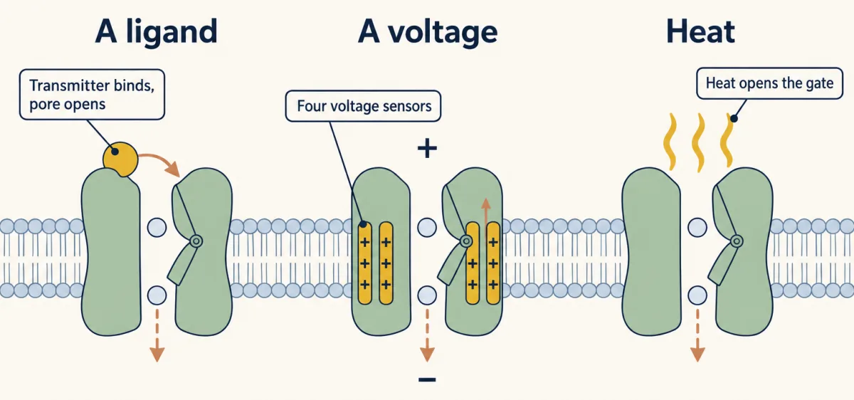
Three examples of channel gating: an extracellular transmitter opens a ligand-gated channel; depolarisation moves the four voltage sensors of a voltage-gated sodium channel; and heat opens a heat-sensitive TRP channel. In each example, the channel itself is the receptor. The inward arrows illustrate ion entry in these examples, not a universal direction of flow. AI-generated illustration -
A channel that needs two things at once, and what plugs a pore
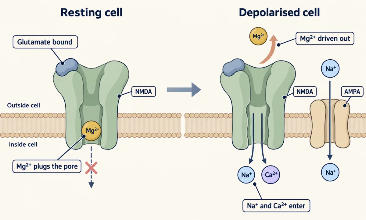
Strong NMDA receptor conduction needs glutamate bound and the cell depolarised, with glycine or D-serine also bound. At rest, extracellular Mg2+ blocks the open pore much of the time, so very little current flows (the X marks a near-complete block, not exactly zero). Depolarisation relieves the block, allowing strong Na+ and Ca2+ entry. Outside the cell is above the membrane in both views. AI-generated illustration 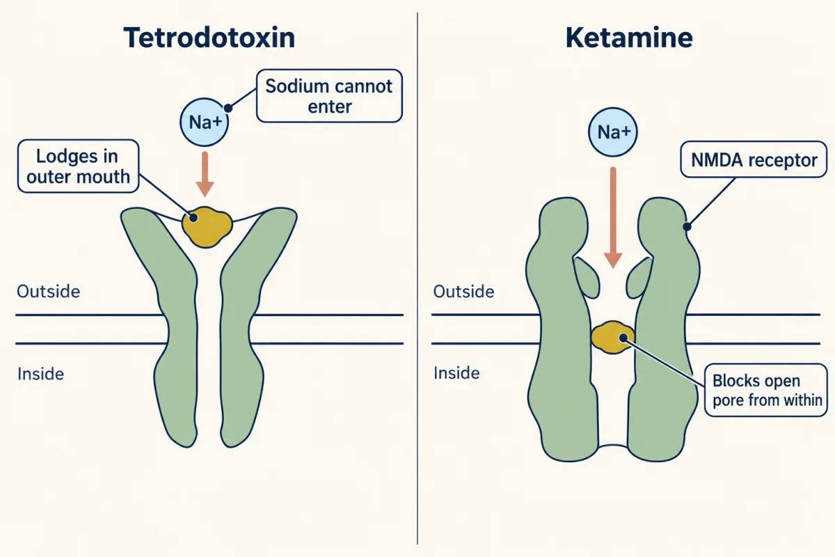
Because a channel is a hole, it can be plugged. Tetrodotoxin lodges in the outer mouth of the voltage-gated sodium channel; ketamine enters the open NMDA receptor pore and blocks it from within. AI-generated illustration
Where this lesson fits
Before thisReview first
- A Field Guide to ReceptorsThis course · Lesson 4
It introduces the acetylcholine-gated muscle channel that this lesson uses as the model of a ligand-gated pore.
- Calcium: The Messenger Kept at Arm's LengthThis course · Lesson 15
Calcium as both an ion and a messenger, with Orai and the IP3 receptor, is assumed when Ca2+ enters through channels here.
- Sensing the World: Vision, Smell, Nitric Oxide and cGMPThis course · Lesson 17
The rod's cGMP-gated channel is one of the channels opened from inside the cell that this lesson names.
After thisStudy next
- Signaling to the Nucleus: CREB, AP-1 and Immediate Early GenesThis course · Lesson 32
Calcium entering through the NMDA receptor sets off a path to the nucleus and immediate early genes.
- Long-Distance Signaling in NeuronsThis course · Lesson 37
The depolarisation and impulse that channels create here must be carried along an axon to reach the cell body.
Elsewhere on the siteFilms, simulations and method films in other courses
- The length constantSimulation · Neuroscience
Resting voltage here comes from the balance of potassium and sodium gradients, which you need to predict ion direction.
- The Spike and Its PuzzlesFilm · Neuroscience
Voltage-gated sodium and potassium channels open and close in sequence to produce the spike.
- Two Pushes, One HillFilm · Cell Biology
It shows how concentration difference and membrane voltage together set the direction of ion flow.
- Why Capsaicin Feels HotFilm · Neuroscience
Capsaicin opens the heat-sensing TRPV1 channel, the same protein as the chilli and hot-stove example.
Suggested by an AI model from the lessons themselves, then checked. How we use AI