Sensory Systems · Skim version
Sensing the Gut: Enteroendocrine Cells and Nutrient Signals
Read the full lesson → Skim the whole course →
The guiding idea
A mouse with no sweet taste still learns to prefer sugar over a sweetener. The sensor is in its gut, and it is not a neuron. How would you show that?
Cells in the lining of the intestine, which are not neurons, sense chemicals in the gut and pass the signal to sensory nerves.
From the film: How to Prove a Gut Synapse
- Anatomy can show a gut cell contacts a nerve, but it cannot identify the transmitter.
- A fast current in a cocultured vagal neuron tests transmission directly.
- A glutamate-receptor blocker that silences the current, followed by washout recovery, supports glutamate transmission.
- In mice, silencing and activating CCK-labelled gut cells test necessity and sufficiency for a vagal sugar response.
From the film: How the Gut Teaches Preference
- In mice, learned sugar preference can develop without sweet-taste signalling.
- Initial attraction and learned preference are different measurements.
- Intestinal SGLT1 function is needed for the gut sugar signal in these mouse studies.
- Sugar and fat have separable receptor requirements and partly distinct vagal populations.
-
First establish a sensory epithelial cell
-
What each synapse experiment earns
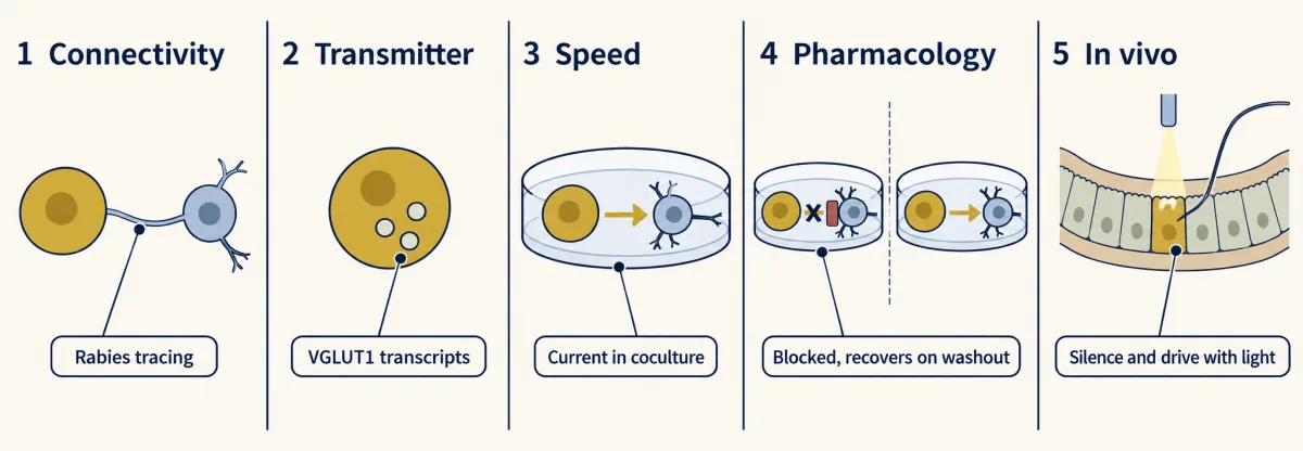
The evidence that an epithelial cell synapses onto the vagus comes in five steps, each answering a different doubt. Each step has a limit the text spells out: for example, connectivity labelling reached vagal neurons in only some mice, and the cells that made contact were a minority. AI-generated illustration 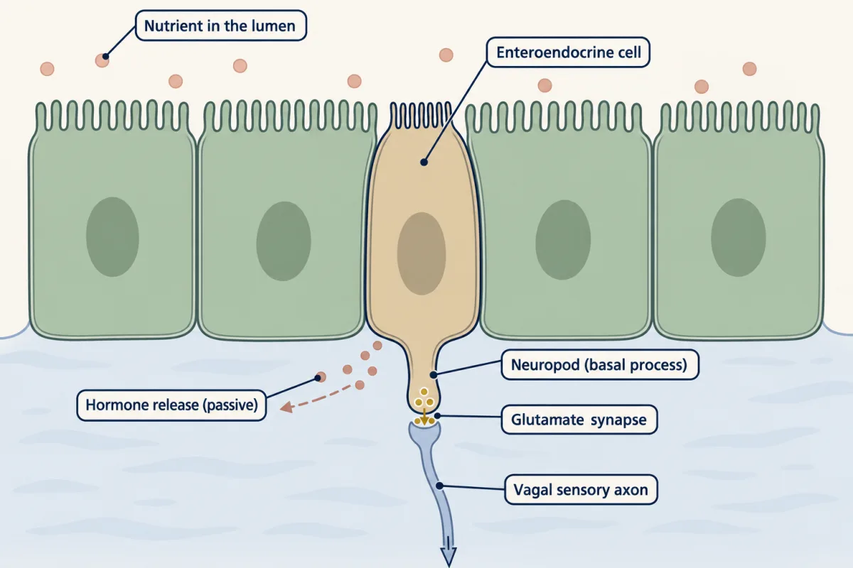
An enteroendocrine cell can signal in two ways. Beside passive hormone release into the tissue, Kaelberer and colleagues (2018) argued for a glutamate synapse from its basal process, the neuropod, onto a vagal sensory nerve. Only a minority of CCK cells were found in contact with sensory fibres in fixed tissue, so this does not describe all enteroendocrine cells. AI-generated illustration -
Sugar without taste: locate each link
-
Sugar and fat can separate
-
Worked problem: preference above chance
-
GLP-1: three routes, not one relay
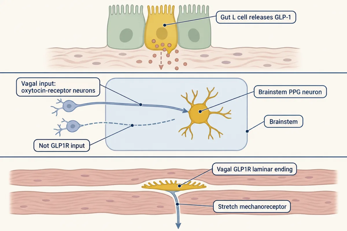
Three different things are called GLP-1 signalling: L cells in the gut lining, preproglucagon neurons in the brainstem, and vagal GLP1R neurons. The rodent evidence does not support a chain from gut GLP-1 through the vagus to brain GLP-1 neurons, so the lanes are kept apart. AI-generated illustration -
Design problem
-
Going deeper
Where this lesson fits
Before thisReview first
- Vagal Sensory Neurons: The Body Reports to the BrainThis course · Lesson 16
This lesson takes the vagal sensory neurons it names and asks which gut cells excite them.
- Five Ways to Turn a Stimulus into a CurrentThis course · Lesson 1
You need the ways a sensory cell turns a stimulus into a current to see how a gut cell does it.
- Sensory Neurons by Their Genes: the Atlas EraThis course · Lesson 3
Reading vagal neuron populations by gene markers, such as VIP or TRPA1 clusters, builds on this atlas approach.
After thisStudy next
- Combining Senses and Predicting ThemThis course · Lesson 29
Combining gut signals with other senses by reliability takes the idea of weighing evidence further.
- Restoring Senses: Implants, Prostheses and Optogenetic VisionThis course · Lesson 30
Restoring or driving sensory pathways by optogenetic activation builds on the stimulation logic used here.
Suggested by an AI model from the lessons themselves, then checked. How we use AI