Biochemistry · Skim version
Gluconeogenesis: Pushing Glucose Back Uphill
Read the full lesson → Skim the whole course →
The guiding idea
When food runs out, your liver (with help from the kidney) builds the glucose your brain and red blood cells still need, and it does it with four new enzymes, three compartments, and real ATP and GTP spent to make the climb.
Gluconeogenesis is how the body builds glucose from non-sugar parts, a job that matters most when food runs out, and its single organizing idea is that it is not glycolysis run backward.
-
Where the carbons come from
-
Three cliffs: why the pathway can’t just run backward
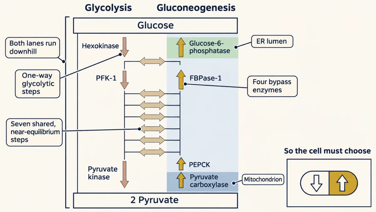
Gluconeogenesis keeps glycolysis's seven near-equilibrium steps and replaces the three one-way steps with four different enzymes (gold), spread over mitochondrion, cytosol and ER. Each lane now runs downhill in its own direction, so thermodynamics no longer decides between them; regulation does. AI-generated illustration -
Bypass 1: pyruvate to PEP, across two compartments
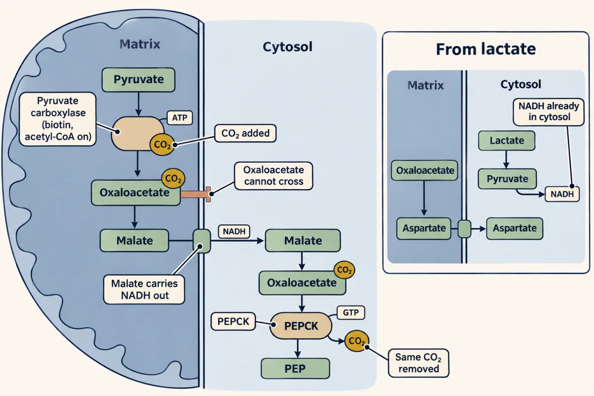
Bypass 1 takes two enzymes and two compartments. Pyruvate carboxylase spends ATP to add CO₂ in the mitochondrion; oxaloacetate leaves as malate, carrying NADH; PEPCK spends GTP and removes the same CO₂ in the cytosol. Starting from lactate, oxaloacetate can leave as aspartate instead, because lactate dehydrogenase has already made the cytosolic NADH. (Human liver also has a mitochondrial PEPCK, not shown.) AI-generated illustration -
Why fat can’t become sugar
-
Bypasses 2 and 3, and why muscle can’t share
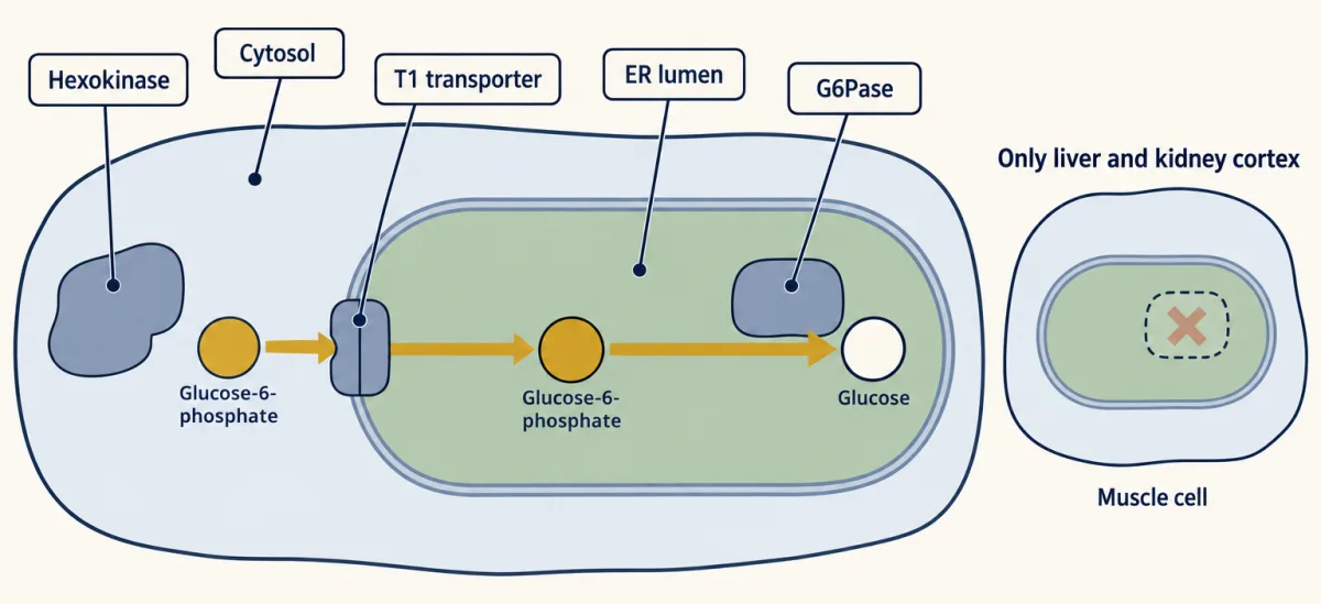
T1 transports glucose-6-phosphate into the ER lumen, where the lumen-facing catalytic site of membrane-bound G6Pase-α releases glucose. Liver, kidney cortex and, to a lesser degree, small intestine express this isoform; skeletal muscle lacks it. G6Pase-β is widely expressed. AI-generated illustration -
The bill
-
Downhill both ways: the switch at each pair
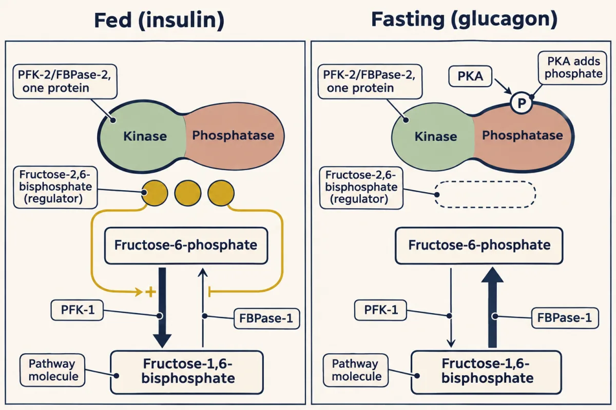
Fed versus fasting at the middle pair. Insulin leaves the bifunctional enzyme dephosphorylated: the kinase makes fructose-2,6-bisphosphate, which activates PFK-1 and inhibits FBPase-1. Glucagon, through PKA, phosphorylates it: the phosphatase destroys fructose-2,6-bisphosphate and FBPase-1 runs. 1,6 is the pathway; 2,6 is the regulator. AI-generated illustration -
When a bypass breaks
-
Going deeper
Where this lesson fits
Before thisReview first
- Glycolysis: The Investment-and-Payoff PathwayThis course · Lesson 16
You need the ten glycolytic steps and the three one-way ones from there to see which steps get bypassed.
- Metabolism: Basic Concepts and DesignThis course · Lesson 15
The free-energy and coupling ideas from there explain why ATP and GTP spent make the climb run downhill.
- Basic Concepts of Enzyme ActionThis course · Lesson 7
You need the idea that an enzyme speeds a reaction both ways but does not set its direction.
After thisStudy next
- The Pyruvate Dehydrogenase Complex: The One-Way Gate to the CycleThis course · Lesson 18
It follows pyruvate the other way, through a one-way step that is why fat cannot become glucose.
- Integration of Metabolism: How the Body Switches FuelsThis course · Lesson 30
It shows how hormones switch the liver between glycolysis and gluconeogenesis as fed turns to fasting.
- Fatty Acid Degradation: The β-Oxidation SpiralThis course · Lesson 25
It shows the fat burning that fuels the liver during a fast and supplies acetyl-CoA to switch on pyruvate carboxylase.
Elsewhere on the siteFilms, simulations and method films in other courses
- What the pump rate measuresSimulation · Physiology
It shows how insulin and glucagon steer liver glucose output, which gluconeogenesis supplies during fasting.
- A Beta Cell Burns Glucose to Read ItFilm · Physiology
It shows how beta cells sense glucose, the signal that decides whether the liver makes or stores glucose.
Suggested by an AI model from the lessons themselves, then checked. How we use AI