Cell Biology · Skim version
Experiments That Distinguish Mechanisms
Read the full lesson → Skim the whole course →
The guiding idea
Where does a protein live, in what order does it travel, and does it cause the defect you see? Each question is best answered by a different experiment, and a pellet named 'mitochondria' is never only mitochondria.
How do you find out where an enzyme lives, in what order a protein visits the compartments of a cell, and whether a protein causes the defect you see when it is lost?
From the film: Where the Twins Disagree
- A result can choose between two models only where the twins disagree.
- In the lesson's made-up table, enrichment in tube L cannot choose, because both models predict it; matching a marker across all five tubes is stronger evidence, though not proof.
- Tagging one batch of protein (a pulse-chase) gives the order, ER then Golgi then granules; carriers and maturing Golgi compartments predict the same order, so the order alone cannot choose.
- In these cancer-cell assays, removing MELK barely moved the drug's growth curve, so its effect there mostly runs through other targets.
- RNAi can silence other messages too, so rescue with an RNAi-resistant copy is strong evidence the intended protein caused the effect.
From the film: Two Jobs, One Protein
- A rescue with a resistant copy strongly supports that the defect came from losing the protein, not which of its jobs mattered.
- Royle and colleagues found in 2005 that silencing clathrin in human cells made mitosis take longer.
- Full-length resistant clathrin rescued mitosis, as both models predict, so that rescue cannot choose between them.
- A failed rescue means little until the construct is shown to be made at about the normal level, folded and correctly placed.
- A separation-of-function mutant keeps one job and loses the other, so the two models can predict different results.
-
Where: breaking the cell carefully
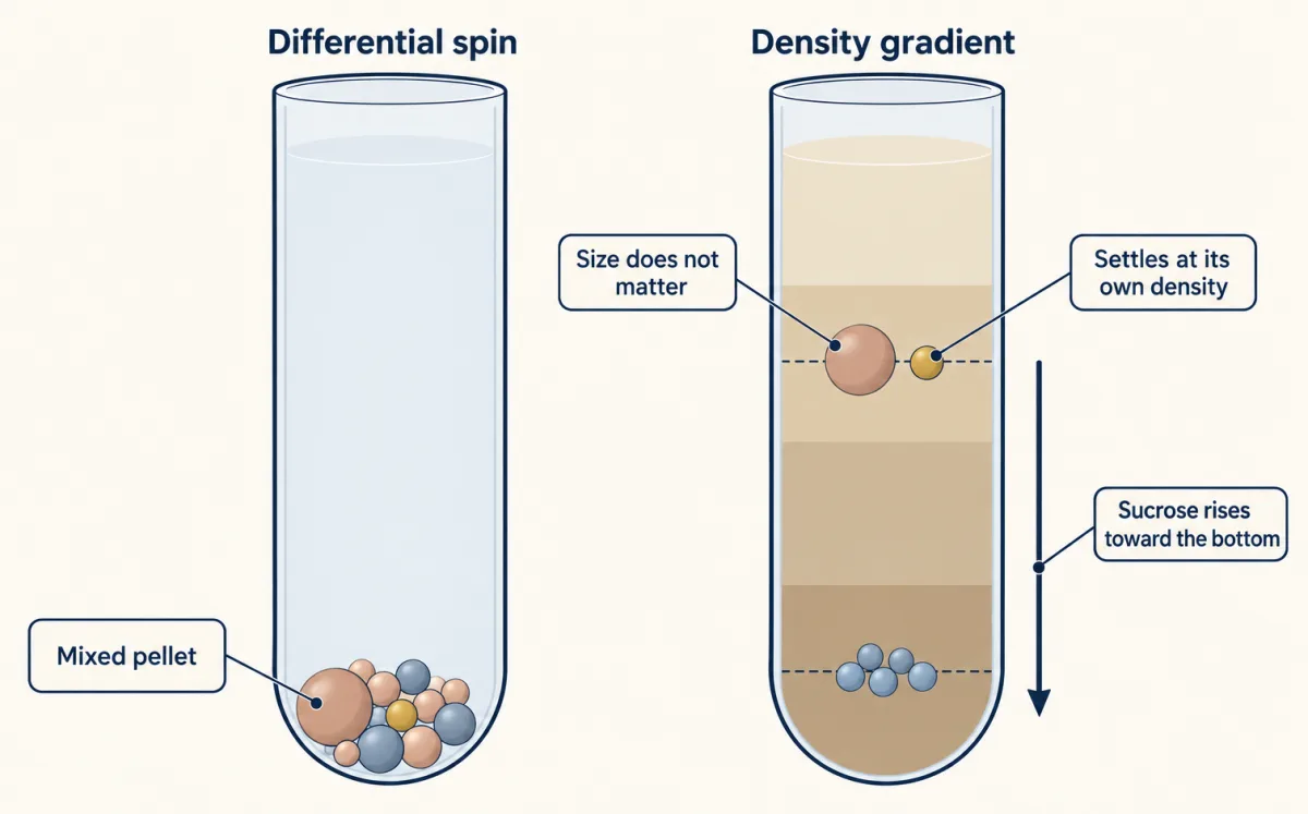
A differential spin gives a mixed pellet; in a sucrose gradient each particle, given long enough, settles at its own buoyant density whatever its size. AI-generated illustration -
When: pulse and chase
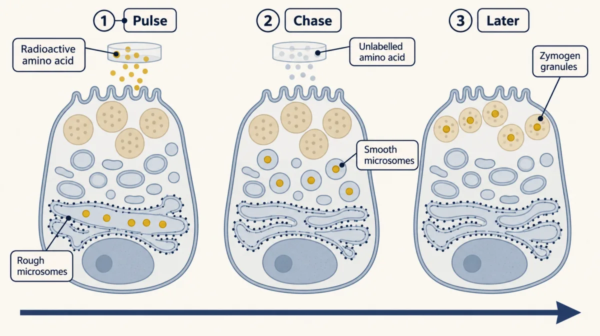
A short pulse tags one batch of protein; after the chase begins, where that batch sits tells you the order of compartments, though the order alone does not show how it moves between them. AI-generated illustration -
Cause: remove it, then put it back
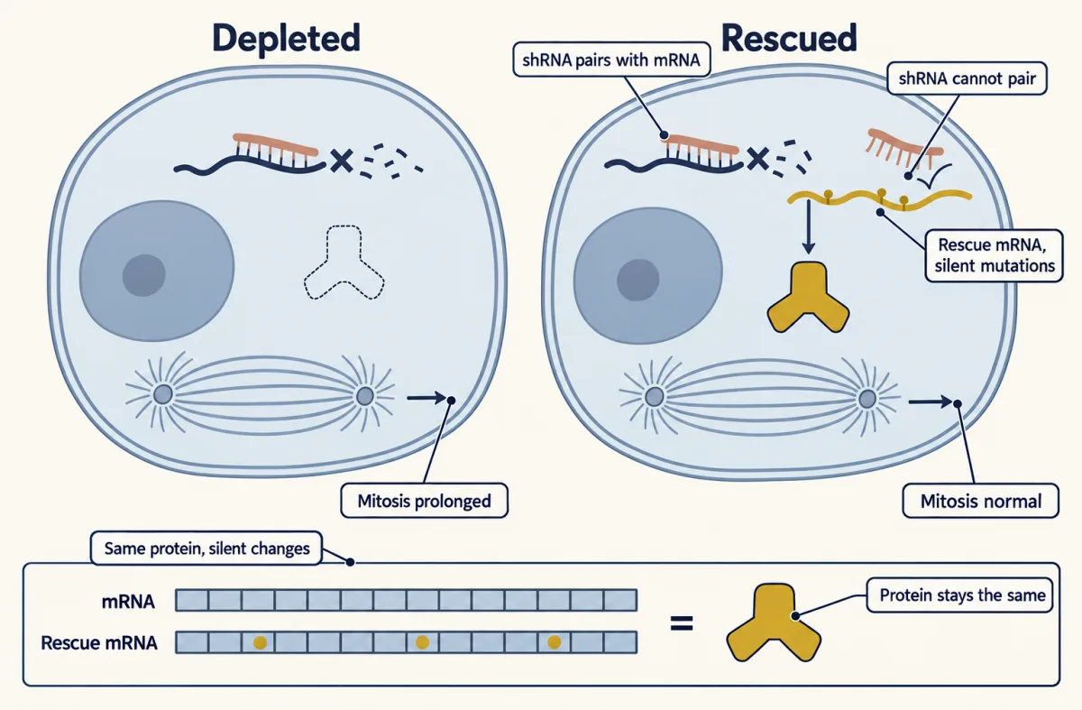
Silent mutations change the mRNA but not the protein, so the shRNA cannot pair with it. If the rescue construct restores normal mitosis, the defect most likely came from losing clathrin. AI-generated illustration -
Going deeper
Where this lesson fits
Before thisReview first
- Microscopy and Quantitative ImagesThis course · Lesson 2
You need its reading of images to follow the autoradiography and the imaging used to confirm where an enzyme sits.
After thisStudy next
- Protein Sorting into CompartmentsThis course · Lesson 7
It uses protection and pulse-chase style evidence to show which compartment a protein reaches and when.
- The Secretory PathwayThis course · Lesson 12
It takes the pulse-chase order of ER, Golgi and granules further and tests carrier against maturation models.
Elsewhere on the siteFilms, simulations and method films in other courses
- Fluctuation test: induced or already there?Simulation · Genetics and Molecular Biology
This fluctuation test is another experiment designed so two models predict different results.
- When Two Mutants Rescue Each OtherFilm · Genetics and Molecular Biology
It shows a test built to separate two explanations of the same mutant phenotype.
- What the Halfway Band MeansFilm · Genetics and Molecular Biology
The density-label experiment is a classic design where competing models predict different outcomes.
Suggested by an AI model from the lessons themselves, then checked. How we use AI