Helices, Sheets and the Rule-Breakers
Read the transcript of “Helices, Sheets and the Rule-Breakers”
Film transcript: Helices, Sheets and the Rule-Breakers
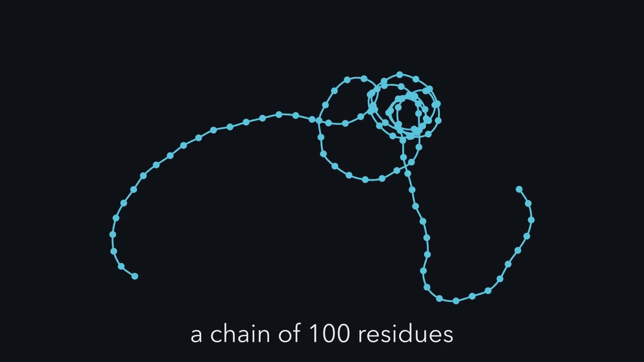
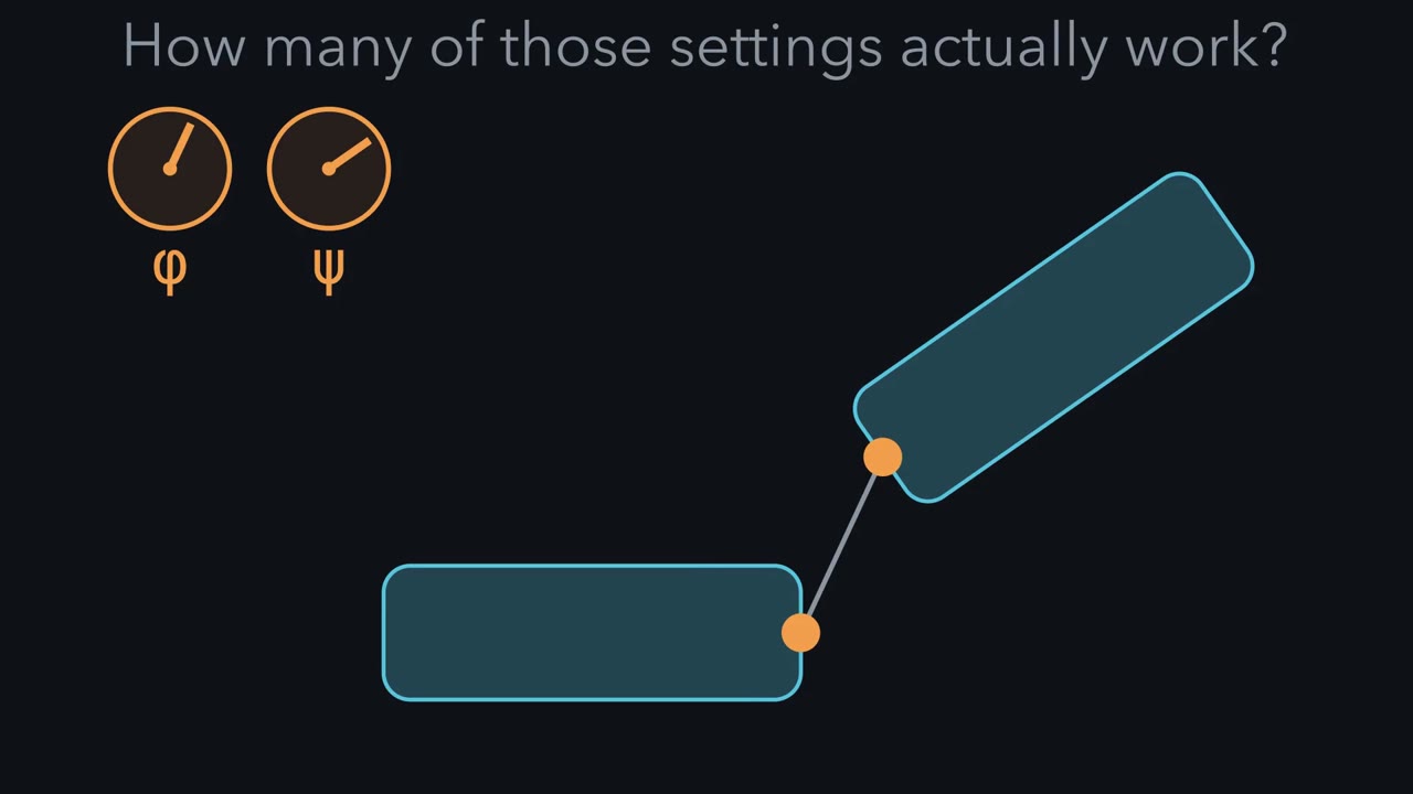
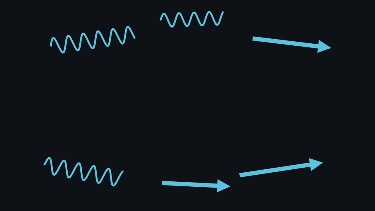
The map has islands. Why do they become helices and sheets? Each island is the geometry of a regular shape. That shape lets the backbone hydrogen bond to itself. The bonds run between backbone C=O groups and N–H groups. Shapes built this way are called secondary structure. Secondary structure is local. It describes a stretch of chain, not the whole protein. First, the alpha helix. It is a right-handed coil. It has a little, over three and a half residues per turn. Inside the helix, each C=O group bonds to the N–H group four residues ahead. In the first turn, the N–H groups have no partner four residues back. The side chains stick outward. In a helix, the partners are near each other in the sequence.
Second, the beta sheet. Its strands are stretched nearly flat. Side chains alternate, above and below the sheet. Each strand bonds to its neighbouring strands. They can run in the same direction, or in opposite directions. Those strands can lie far apart in the sequence. Now meet a rule-breaker, proline. Its side chain loops back and bonds to its own backbone nitrogen. That makes a ring. So proline has no N–H group to donate, and its ring is bulky. Here is the test. Where in a helix could a proline sit without breaking it?
In the first turn. There, the N–H groups have no partners anyway. Losing one costs nothing. In the middle, proline breaks the helix. It cannot donate its hydrogen bond. And its ring bumps into the backbone of the turn before it. Its fixed angle is not the problem. That angle is a helical one. The same ring builds something else, collagen. It pre-shapes each strand into an extended helix. Collagen repeats a pattern: Gly-X-Y. X and Y are often proline and hydroxyproline. Three chains wind together into a triple helix. Every third residue sits at the crowded center. Only glycine is small enough to fit there. Put a larger residue in place of that glycine, in type I collagen.
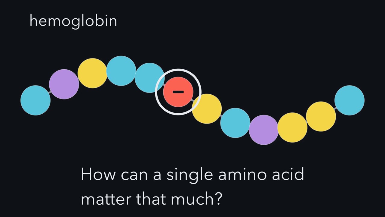
That is a common cause of osteogenesis imperfecta, brittle-bone disease. What kills one structure builds another. One residue, again, decides the shape. Geometry allows these local shapes. But it does not choose which ones a protein makes. Helices and sheets are the backbone bonding to itself.
One sequence, one working shape. Change a single letter and the shape, and the job, can break.
Continue in the lesson: Protein Three-Dimensional Structure: The Sequence Sets the Shape →
2:58 · Free, with captions and a transcript. The lesson has the full text, the questions and the sources.
More films from this lesson
How this film was made. Narration: an AI narrator voice (Kokoro, af_heart) reading narration written from this lesson's text. Animation: AI-generated in code (Manim) by Claude, a schematic with no drawn molecular structures. Captions come from the narration script. See something wrong? Report it.