Films · Developmental Neuroscience
One Cell, Two Eyes
Read the transcript of “One Cell, Two Eyes”
Film transcript: One Cell, Two Eyes
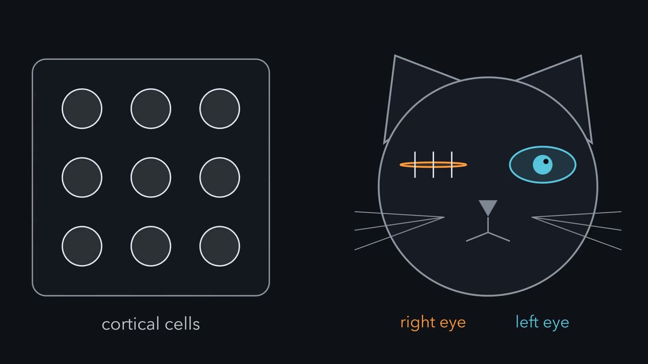
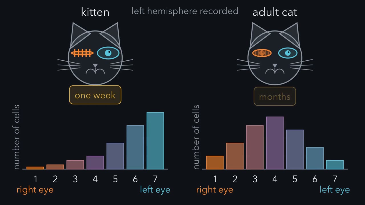
Close one eye of a young kitten for a week, and its visual cortex changes. Close an adult cat's eye for months, and the cortex looks normal. Why? To see what changed, follow the signal from the eyes to the cortex. Each eye sends its axons to a relay station in the thalamus, the lateral geniculate nucleus. We will call it the LGN. There the two eyes' axons end in separate layers. So most LGN cells are driven by one eye. LGN axons carry the signal on to the primary visual cortex, at the back of the brain. They end mainly in layer 4. There the two eyes' endings still lie apart, in alternating stripes. In macaque monkeys these stripes, called ocular dominance columns, are sharp.
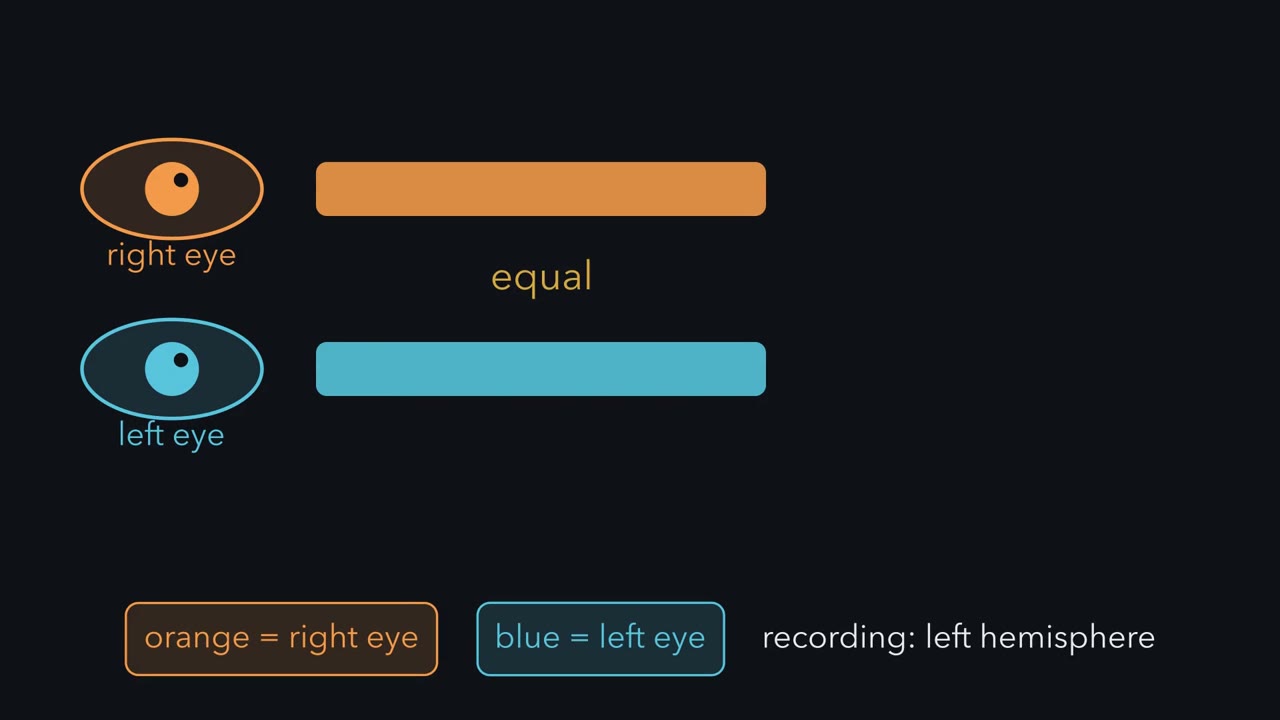
In cats the pattern is less regular. In macaque layer 4C, most cells are driven mainly by one eye. Cat layer 4 has more cells that hear both. Above and below layer 4, cortical neurons combine the two streams. Many cells there can be driven by both eyes. In cats and monkeys, this is the first place where many single cells are driven by both eyes. The eyes compete for territory in layer 4, and for those shared cells. Which eye is which depends on where you record. We will always record from the left hemisphere. The eye on the opposite side, the right eye, is contralateral, in orange. The left eye, on the same side, is ipsilateral, in blue. David Hubel and Torsten Wiesel measured the balance one cell at a time.
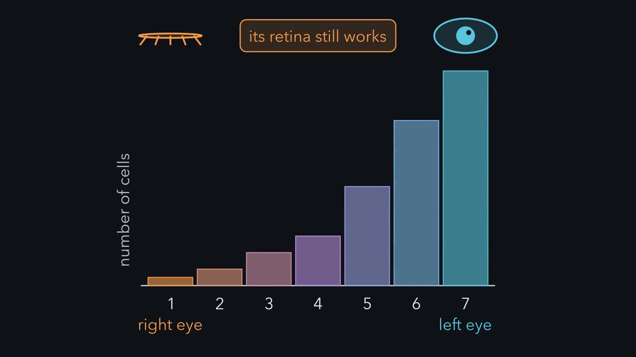
A fine metal electrode sits next to one neuron and records its spikes from outside the cell. With the animal anaesthetized, they showed a bar of light to one eye, with the other covered. Then they switched. Each cell went into one of seven ocular dominance groups. Group 1 answers only to the contralateral eye. Group 7 answers only to the ipsilateral eye. Group 4 answers about equally to both. The groups between favor one eye, more or less. One worked cell fires to the right eye and not at all to the left. That puts it in group 1. A cell that fires only to the left eye goes in group 7. A cell no eye can drive is counted as unresponsive. Count the cells in each group, and you get an ocular dominance histogram.
In a normal adult cat it is a hump. Most cells sit in the middle groups, leaning toward the contralateral side. Takeaway: the eyes' paths stay apart until the cortex, where shared cells can be driven by both. So what sets each cell's group? The next chapter closes one eye and watches the balance move.
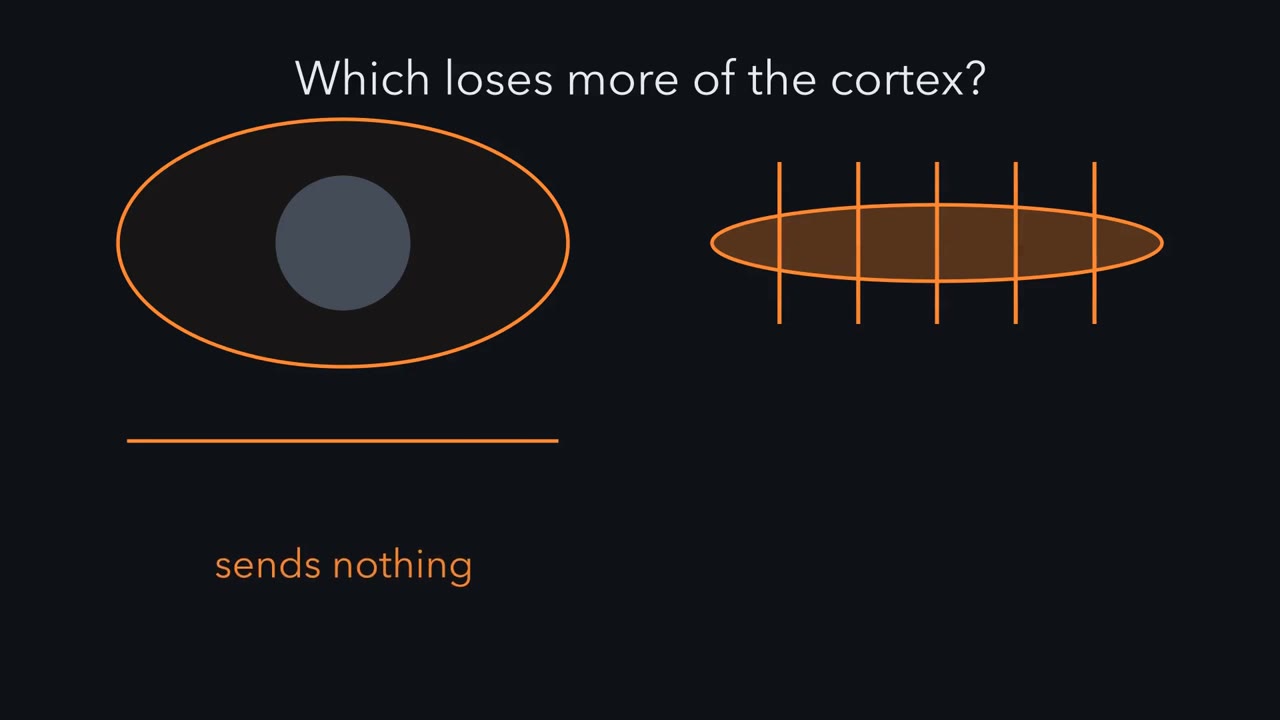
Why does closing one eye of a kitten for a week rewire its cortex, when closing both eyes shifts the balance much less?
Continue in the lesson: Ocular Dominance and the Critical Period →
3:03 · Free, with captions and a transcript. The lesson has the full text, the questions and the sources.
More films from this lesson
How this film was made. Narration: an AI narrator voice (Kokoro, af_heart) reading narration written from this lesson's text. Animation: AI-generated in code (Manim) by Claude, a schematic with no drawn molecular structures. Captions come from the narration script. See something wrong? Report it.