Two Inputs, One Exit
Read the transcript of “Two Inputs, One Exit”
Film transcript: Two Inputs, One Exit
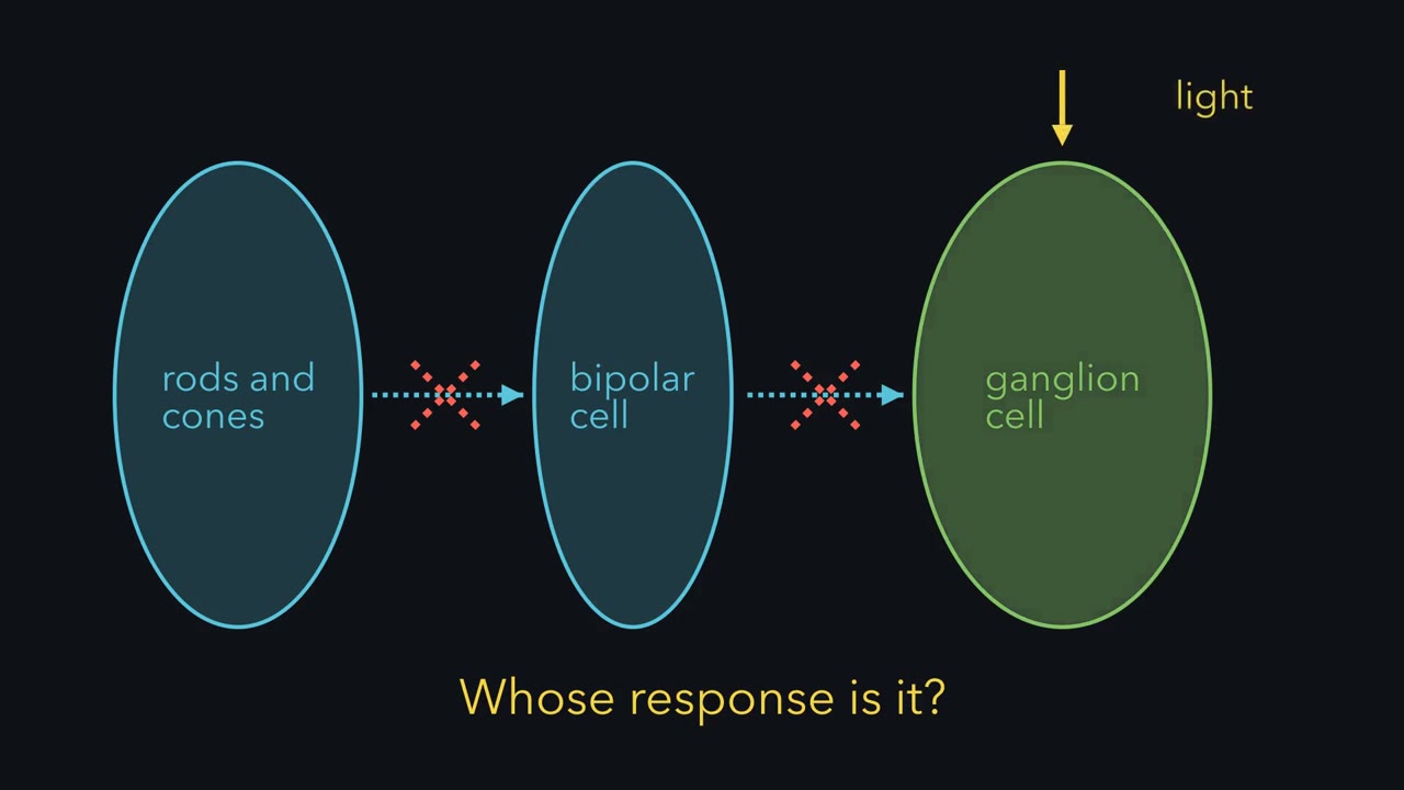
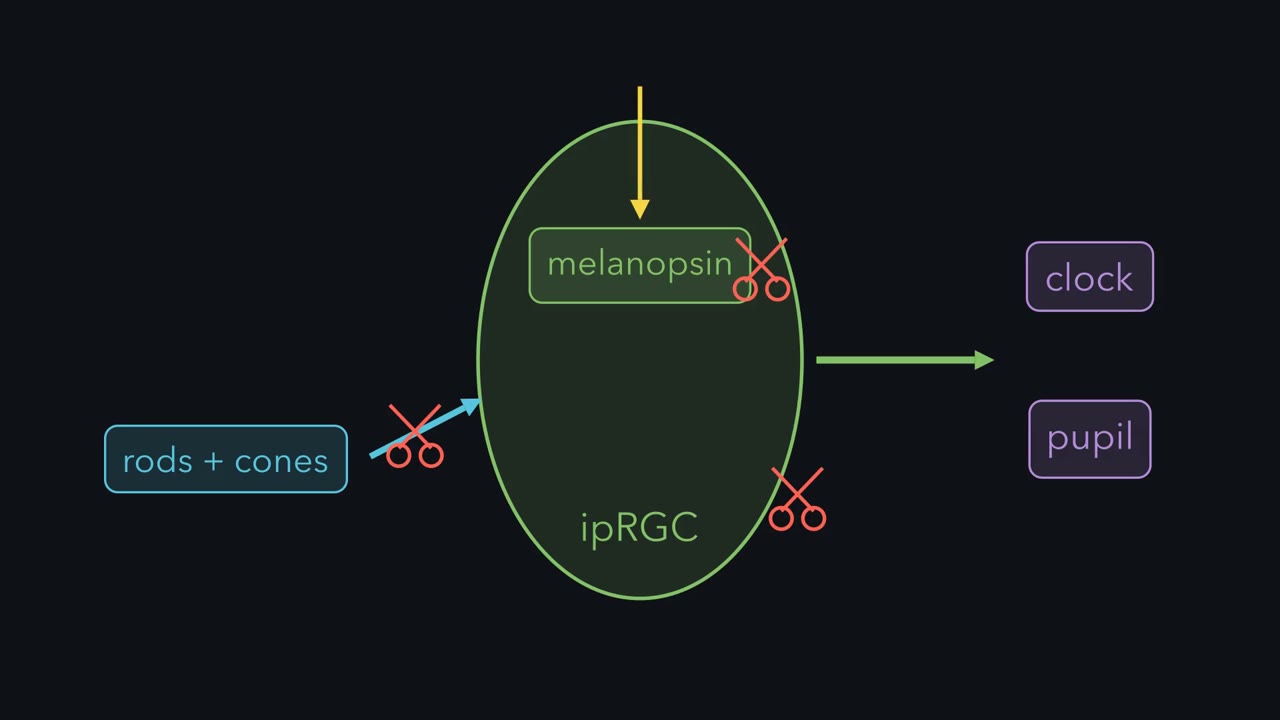
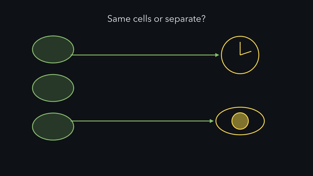
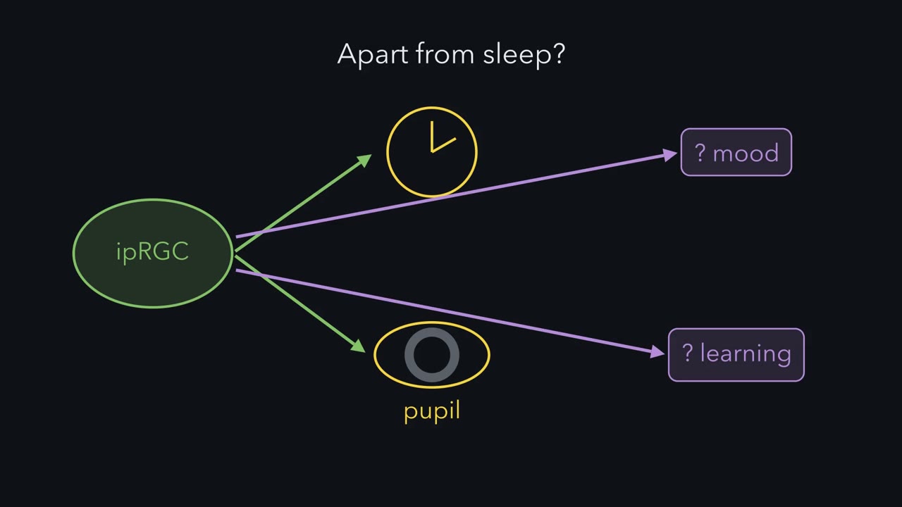
Light hyperpolarizes a rod. Yet light depolarizes these ganglion cells. How can the same light push two cells opposite ways? Start with the wiring. Rods and cones signal to bipolar cells. Bipolar cells signal to the ganglion cell's dendrites. Melanopsin sits in the ganglion cell's own membrane. So light reaches the cell by two routes. But each cell has one axon, one exit. Across the population, those exits reach the clock and the pupil centre. That is the one idea of this lesson. An ipRGC pools two light signals into one output. One input is the rod and cone relay, usually faster and more sensitive. The other is melanopsin, usually slower and less sensitive.
Both can be sustained, and both adapt. In mice, these cells are the main route by which either signal reaches the clock and the pupil. So removing the pigment, or the rods and cones, leaves the other input working alone. Removing the cell removes both. Now the polarity. In a rod, light closes cation channels. Less positive charge enters, and the rod hyperpolarizes. In an M1 cell, light opens channels instead. Melanopsin is an opsin. But it is closer to the visual opsins of insects than to rod and cone opsins. It activates a G protein, inferred to be of the Gq family. That activates phospholipase C beta 4. Then TRPC6 and TRPC7 channels open. Without phospholipase C beta 4, the M1 cell's own light response essentially disappeared.
A fly photoreceptor works much the same way, with Gq, phospholipase C and TRP channels. Predict. These channels pass cations, and their current reverses near zero millivolts. Opening them moves the voltage which way? Recall the balance-point rule. When the voltage is steady, with no injected current, it sits at a weighted average of reversal potentials. The weights are the conductances. Here that is near zero. So the voltage moves up, away from the potassium potential. The cell depolarizes. In Another subtype, M4, may also been reported to close potassium leak channels. That depolarizes too, by the same rule. Which other channels also contribute in M4 is still disputed.
Light hyperpolarizes a rod and depolarizes an M1 cell. And both of the cell's inputs leave by one exit.
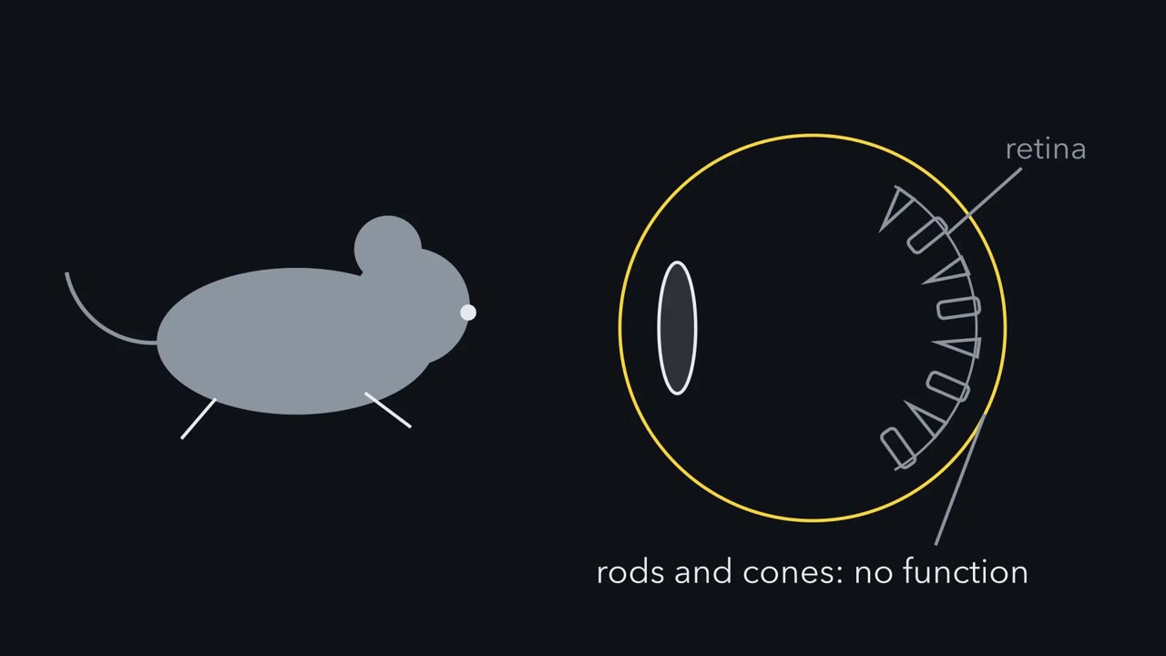
A mouse with no working rods or cones still sets its body clock to the day and still narrows its pupils in bright light. Which cells are measuring the light?
Continue in the lesson: Melanopsin Cells: Light for the Clock and the Pupil →
3:07 · Free, with captions and a transcript. The lesson has the full text, the questions and the sources.
More films from this lesson
How this film was made. Narration: an AI narrator voice (Kokoro, af_heart) reading narration written from this lesson's text. Animation: AI-generated in code (Manim) by Claude, a schematic with no drawn molecular structures. Captions come from the narration script. See something wrong? Report it.