The Light Response That Survived the Block
Read the transcript of “The Light Response That Survived the Block”
Film transcript: The Light Response That Survived the Block
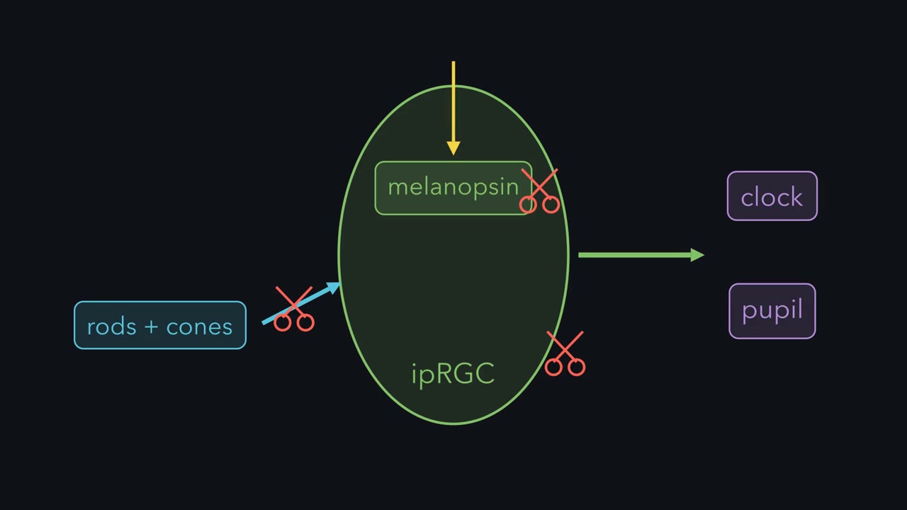
Cut a ganglion cell off from rods and cones. If it still responds to light, whose response is it? Here is the logic. Rods and cones reach ganglion cells through synapses. Block those synapses with drugs. If a ganglion cell still responds once the block is verified, the response is most likely its own. In 2002, Berson, Dunn and Takao did this in rat retina. First they labelled ganglion cells from the SCN. That told them which cells project to the clock. Then they recorded from those labelled cells, while the blockers were applied. Predict. Suppose the rod and cone relay were the cell's only light signal. What should the recording show after the block?
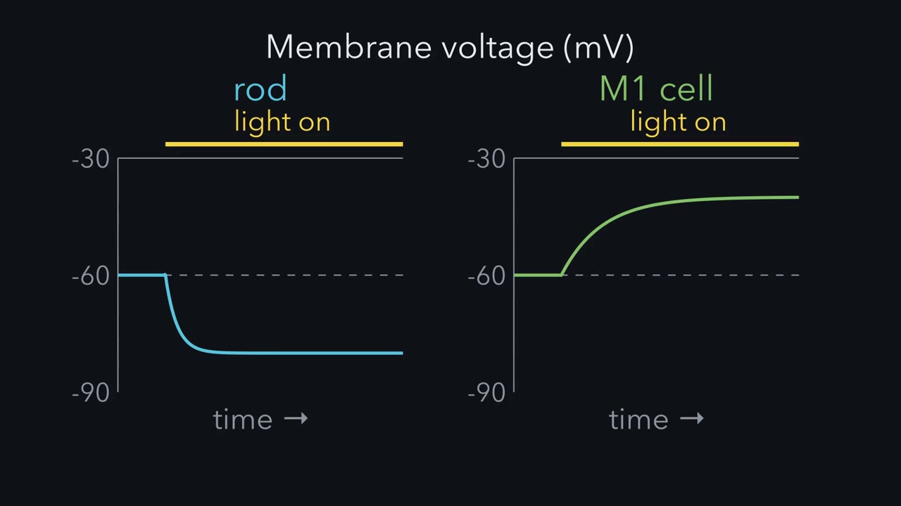
There should be no light-evoked change. But one remained. The labelled cells still depolarized to light. So the response was intrinsic, made within the ganglion cell itself. They also recorded from a cell body removed from the retina. That is a stronger test, with no synapses left at all. So one cell can give two kinds of light response. Put both on one time axis. The relayed response, from rods and cones, usually starts fast and works in dim light. It can have both brief and lasting parts. The cell’s own response is usually slow to start and needs brighter light. It is often long-lasting, though it adapts too. When the light goes off, it decays slowly.
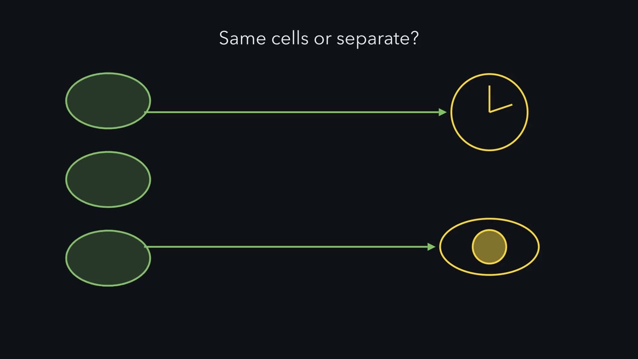
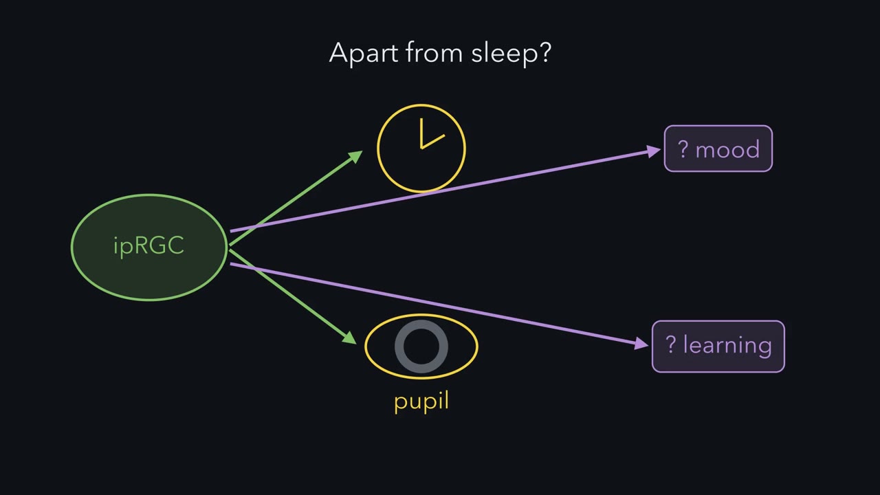
The cell stays depolarized for a while. In people, these cells drive the post-illumination pupil response. The pupil stays small for a while after light goes off. Why would slow and sustained be useful? The clock and the pupil need to know how bright it is. A camera must follow fast changes. A light meter needs a steady reading. The cells that serve the clock and the pupil. are mostly a type called M1. They report how bright the world is, not what is in it. The rat response fitted a pigment that peaks near 484 nanometres. That is a rat estimate, not a human one. In the same volume of the journal, Hattar and colleagues showed that these rat cells carried melanopsin.
And melanopsin-marked mouse axons reached the SCN. A response, a pigment and a target now pointed to one set of cells. Their signature is slow, insensitive and sustained.
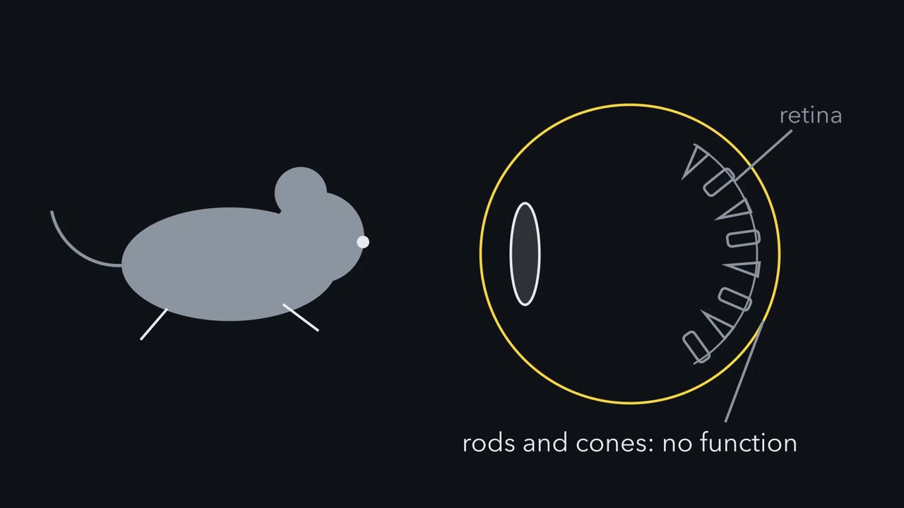
A mouse with no working rods or cones still sets its body clock to the day and still narrows its pupils in bright light. Which cells are measuring the light?
Continue in the lesson: Melanopsin Cells: Light for the Clock and the Pupil →
2:54 · Free, with captions and a transcript. The lesson has the full text, the questions and the sources.
More films from this lesson
How this film was made. Narration: an AI narrator voice (Kokoro, af_heart) reading narration written from this lesson's text. Animation: AI-generated in code (Manim) by Claude, a schematic with no drawn molecular structures. Captions come from the narration script. See something wrong? Report it.