Remove the Pigment, or Remove the Cell
Read the transcript of “Remove the Pigment, or Remove the Cell”
Film transcript: Remove the Pigment, or Remove the Cell
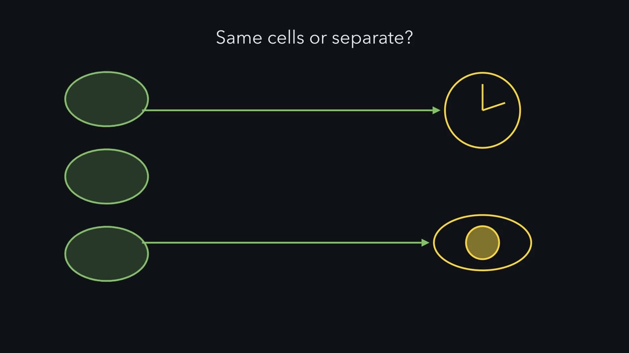
To learn what a part is for, take it away. But which part should you take away? The one idea gives three choices. Remove the pigment. Remove the rods and cones. Or remove the cell. First, the readouts. For the clock, keep a mouse on a light-dark cycle and watch its running. A mouse entrains if its running keeps a fixed timing relative to the cycle. A mouse whose clock is not entrained free-runs. It drifts on its own internal period. Separately, a brief light pulse at night tests how far the rhythm phase-shifts. Masking is a third readout. Bright light at night often suppresses a mouse’s running right away, independently of any clock shift. The same light can also shift the clock.
For the pupil, film the constriction over seconds to minutes of steady light. Row one is wild type. It entrains, shifts fully, and its pupil constricts fast and holds. Row two has no melanopsin. It still entrains, but its phase shifts to brief pulses are smaller. Its pupil constricts, but in steady bright light the constriction fades over minutes. Row three has no rod or cone signalling. It entrains. Its pupil is slower and less sensitive, but it holds. In bright light, it reaches its normal maximum. Row four is the triple mutant. It lacks melanopsin, rod transducin and a cone channel subunit. It does not entrain. It free-runs, shows no masking and has no significant pupil reflex.
Read down the table. Either input alone is enough to entrain and to constrict. Lose both, and nothing is left. Predict. Keep the rods and cones working, and ablate ipRGCs. If rods and cones had their own route, what would you see?
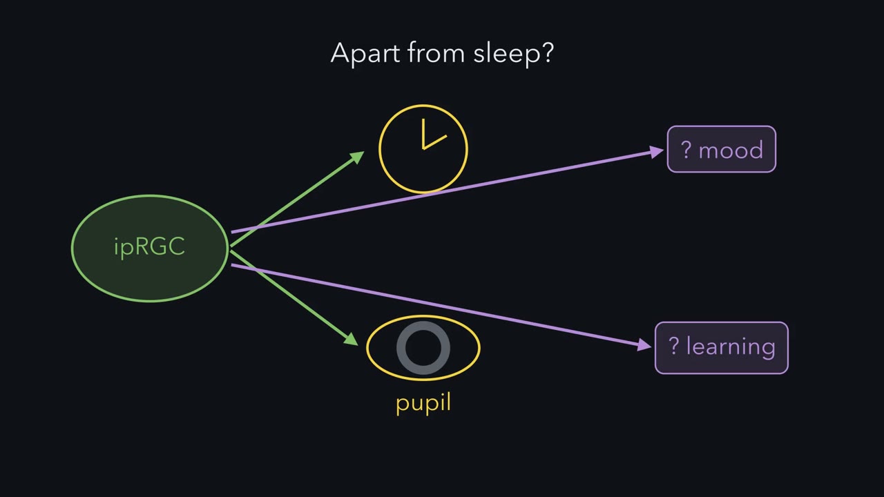
A strong separate route would let these mice keep much of their clock and pupil responses, like the melanopsin knockouts. Güler and colleagues found something else. The deficits were larger than with melanopsin loss alone, similar to those of the triple mutants. Pattern vision, which runs through other ganglion cells, remained. So in mice, these cells are the main route to the clock and the pupil. An ablation removes both inputs, so it cannot say which input supplied what was lost. Predict. Here are made-up phase shifts. Do the two single losses add up to the loss of both?
They do not. Together the two single losses are smaller than the loss when both inputs go. One possible reading is that the output saturates. A pupil cannot get smaller than its minimum. A phase shift has a ceiling too. Near a ceiling, the stronger input alone gets close, and adding the other gains little. A single knockout shows what the other input can do alone. It does not show each input's share. One limit. In the Güler mice, a weakened toxin gene at the melanopsin locus kills the cells gradually and incompletely, and more are lost with age. Losing cells during development can let targets rewire. An ablation timed in adults is better. Remove the pigment, and the relay carries on.
Remove the cell, and both inputs go.
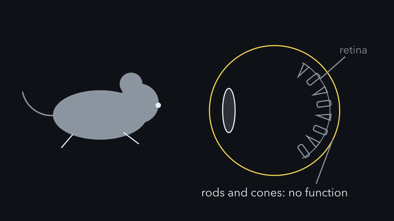
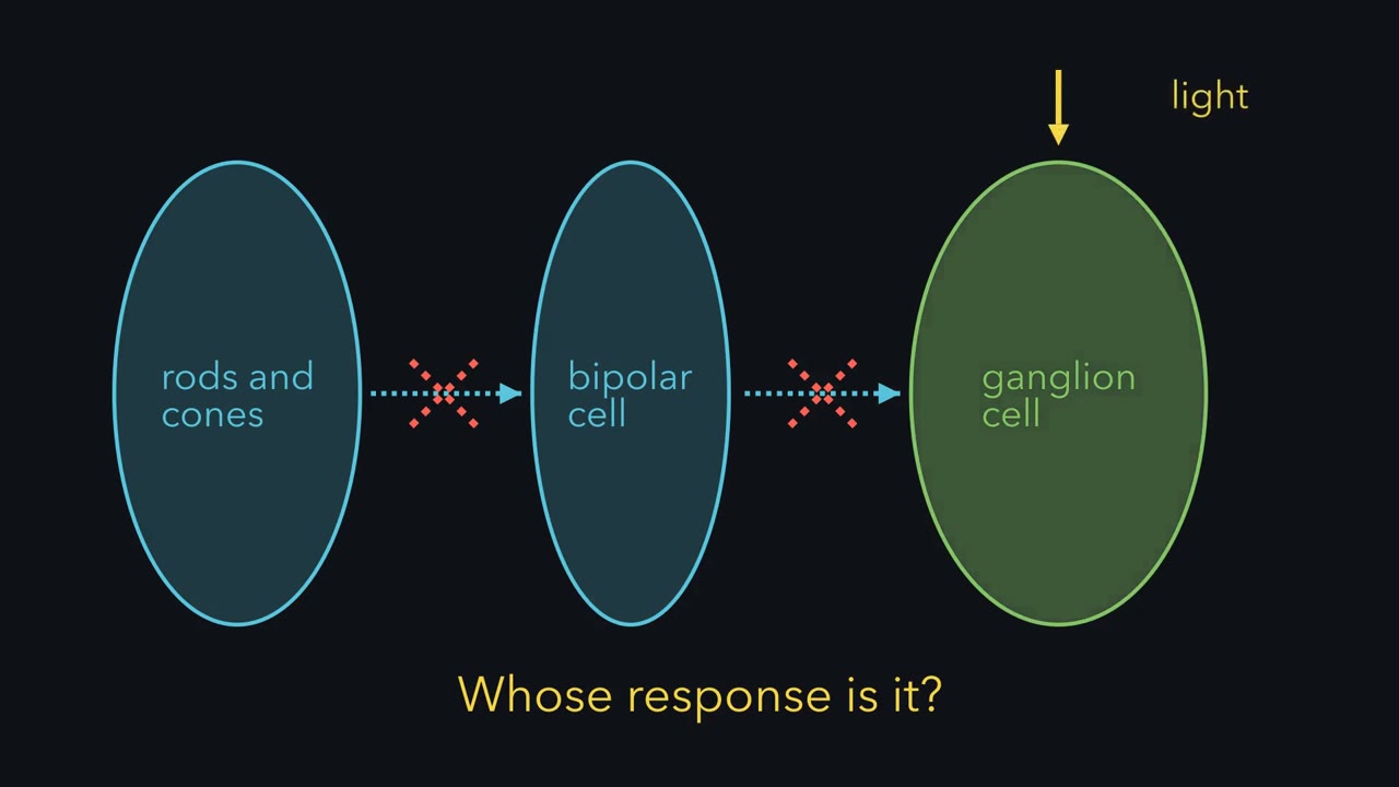
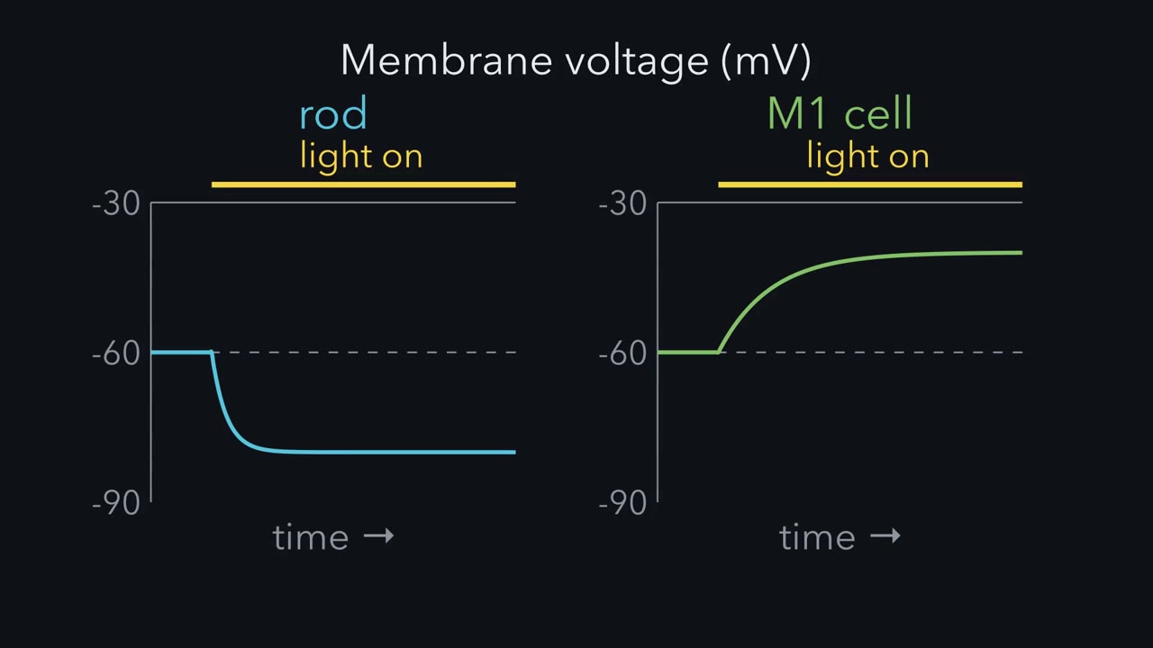
A mouse with no working rods or cones still sets its body clock to the day and still narrows its pupils in bright light. Which cells are measuring the light?
Continue in the lesson: Melanopsin Cells: Light for the Clock and the Pupil →
3:53 · Free, with captions and a transcript. The lesson has the full text, the questions and the sources.
More films from this lesson
How this film was made. Narration: an AI narrator voice (Kokoro, af_heart) reading narration written from this lesson's text. Animation: AI-generated in code (Manim) by Claude, a schematic with no drawn molecular structures. Captions come from the narration script. See something wrong? Report it.