Films · Developmental Neuroscience
Two Answers, a Year Apart
Read the transcript of “Two Answers, a Year Apart”
Film transcript: Two Answers, a Year Apart
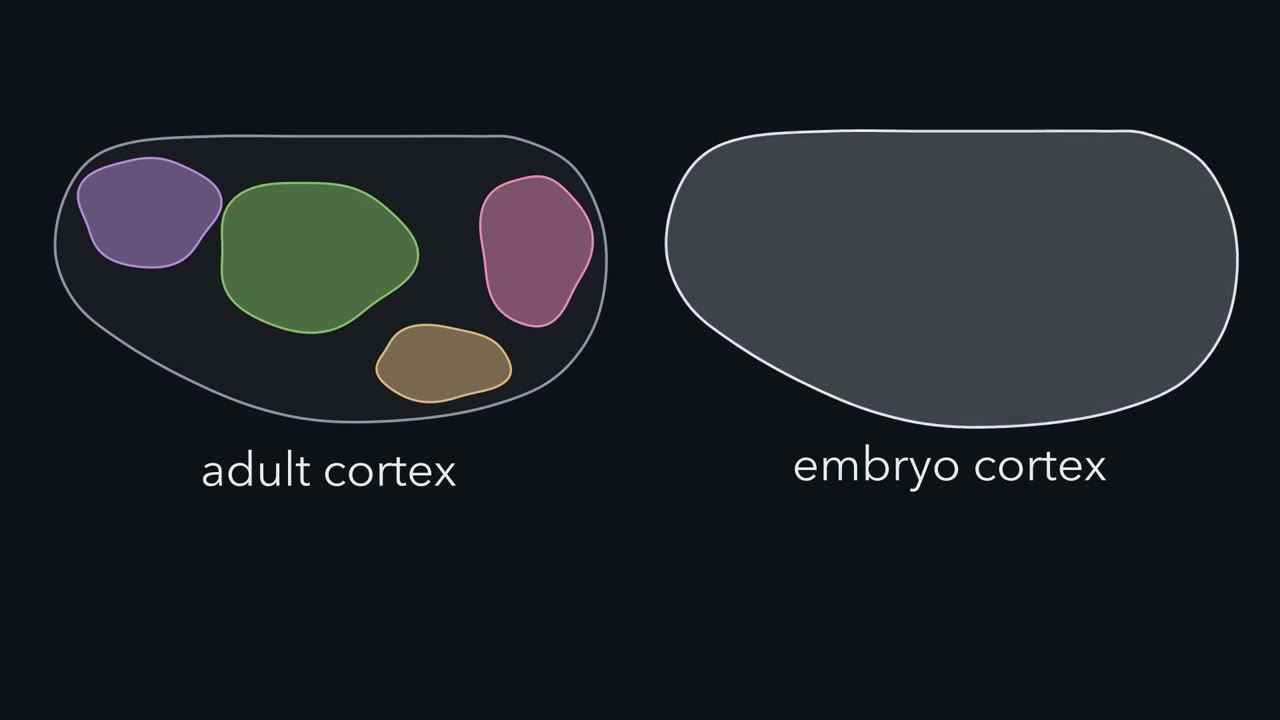
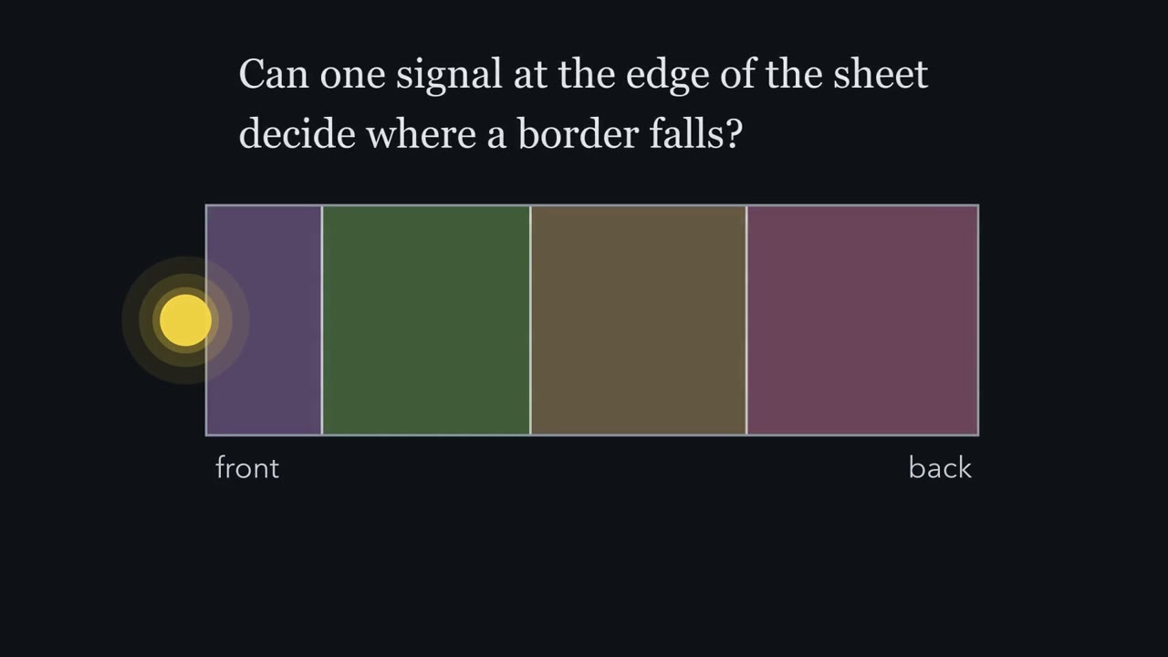
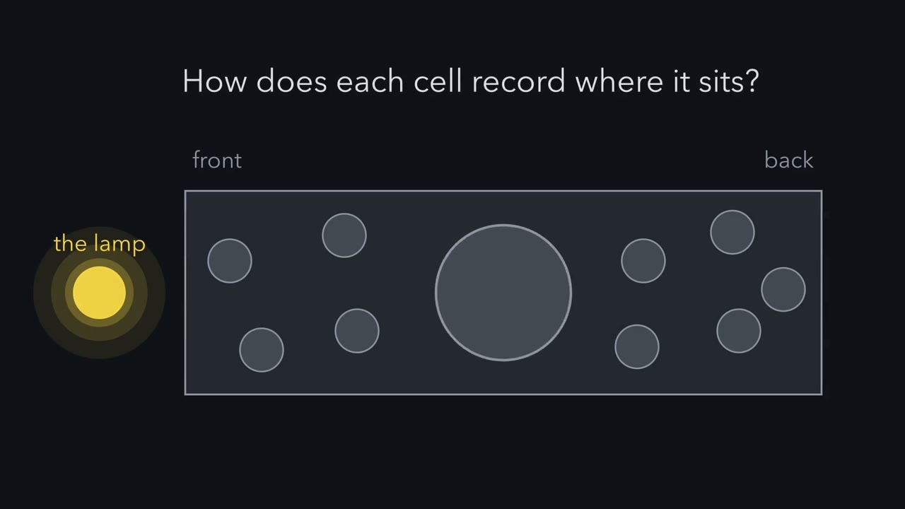
Is the map already in the dividing cells, or is it brought in by the axons that arrive? Two answers were proposed a year apart. In 1988, Pasko Rakic proposed the protomap. Cortical progenitors lie in the ventricular zone, the layer of the brain wall next to the fluid-filled ventricle. Newborn neurons climb outward along radial glia, long scaffold cells that span the wall. So neurons born from one patch stack up in a column above it. If the patches already differ by position, the outline of the areas is laid down where the cells are made. Think of a city zoned into districts before a single house is built. In 1989, Dennis O'Leary proposed the protocortex.
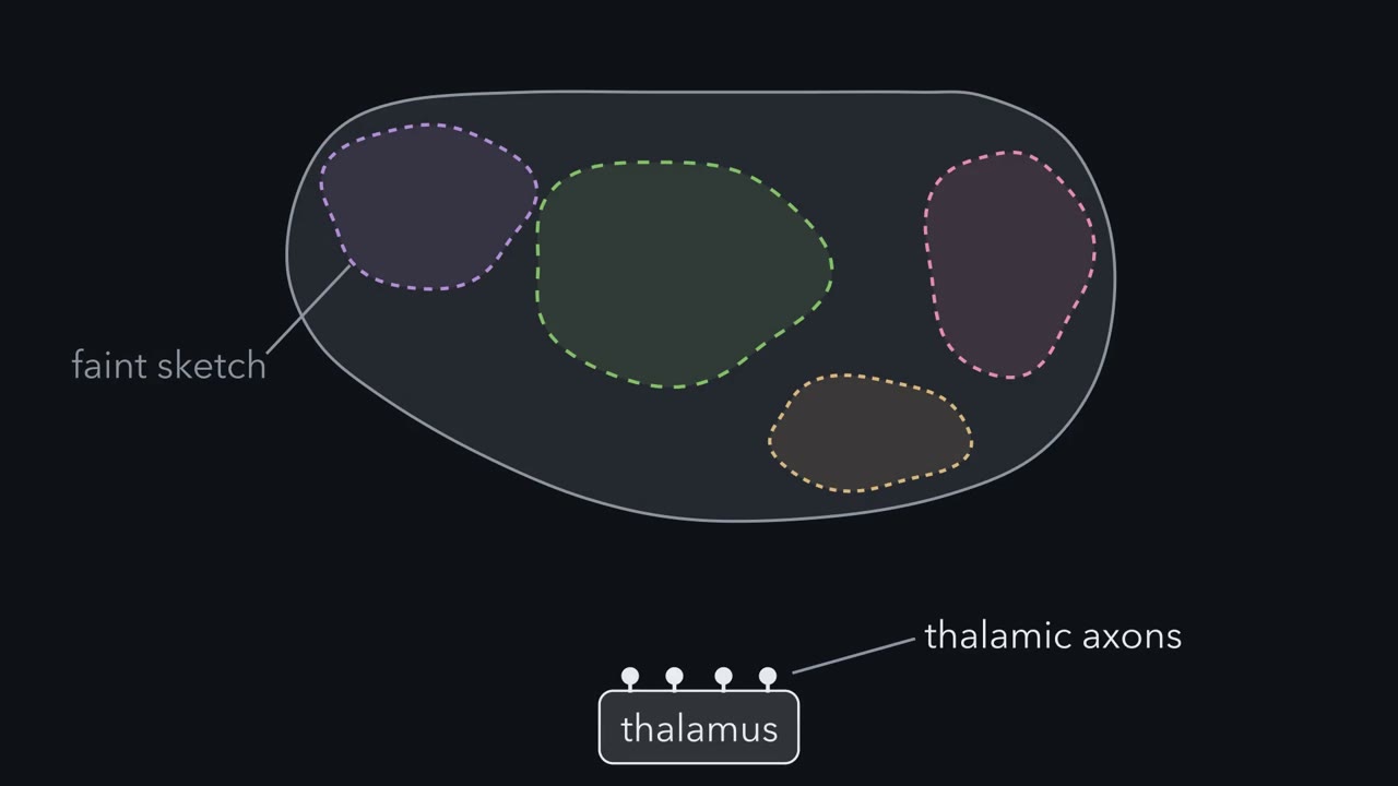
Here the young sheet has broadly similar potential everywhere. Areas emerge mainly from the axons that arrive from the thalamus. Different thalamic nuclei send axons to different parts of the sheet. The input tells each part what to become. Suppose you found molecular differences across the progenitors, and a map that forms without thalamic input. Which idea would that support?
The protomap. That is what it predicts. The protocortex predicts something else. Young cortex given new input should take on features that match the input. Both predictions came true in part. So the field now treats the map as drawn twice. First a sketch from the sheet, then a copy refined by its inputs. One more thing before any result: how do you read a map in a newborn mouse? Mice lacking Emx2, and mice lacking working Pax6, die around birth, before the areas mature. So an area in these experiments means a proxy. First proxy: marker genes. A stain shows where a gene is switched on in a tissue section. Some genes have borders at birth that line up with future areas.
Second proxy: connections. A thalamic nucleus is a cluster of neurons. Each one projects mainly to its own part of cortex. Dye placed in one spot of cortex labels the thalamic neurons that project there, sometimes in more than one nucleus. Third proxy: the barrel field. In mice that survive past birth, a stain on flattened cortex shows the barrels as dark patches. In mice that die at birth, these proxies stand in for mature areas. Animals that survive allow later and adult measures too. The takeaway: the two ideas make different predictions, and early proxies are one way to test them.
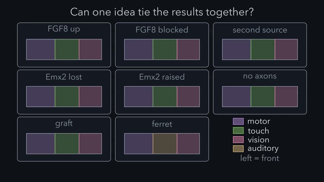
Add one secreted protein to the back of a mouse embryo's cortex and a second, partial whisker map can appear there.
Continue in the lesson: Mapping the Cortex: Areas and Arealization →
3:02 · Free, with captions and a transcript. The lesson has the full text, the questions and the sources.
More films from this lesson
How this film was made. Narration: an AI narrator voice (Kokoro, af_heart) reading narration written from this lesson's text. Animation: AI-generated in code (Manim) by Claude, a schematic with no drawn molecular structures. Captions come from the narration script. See something wrong? Report it.