Films · Developmental Neuroscience
A Sheet with No Borders
Read the transcript of “A Sheet with No Borders”
Film transcript: A Sheet with No Borders
The adult cortex is divided into areas. The embryo's cortex shows no borders at all. So where do the areas come from? The cortex is the sheet of neurons on the surface of the forebrain. In 1909, Korbinian Brodmann divided the human cortex into areas by how its cells were packed and layered. Many of his borders match borders of function. His area 17 is primary visual cortex, the first cortical stop for signals from the eyes. This lesson works in the mouse. Here is the map we will use throughout. Front to back runs from the nose end to the back of the head. The medial edge runs along the midline. The lateral edge is the outer side. Motor cortex sits at the front.
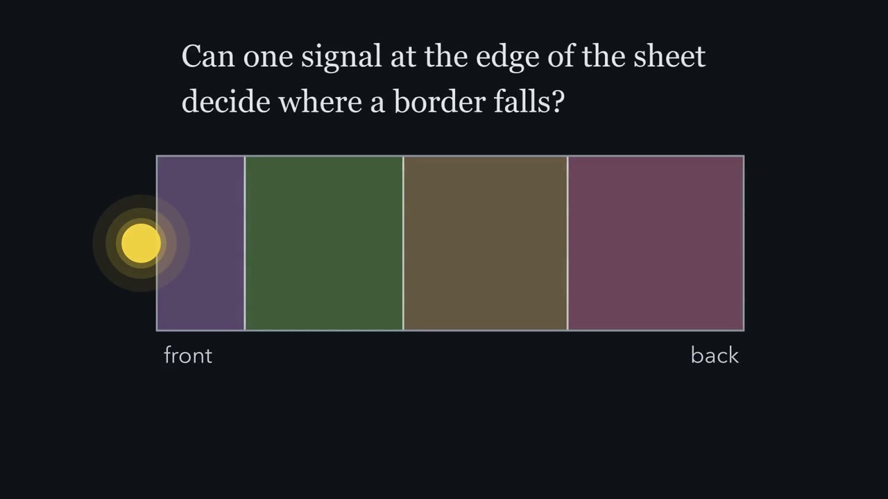
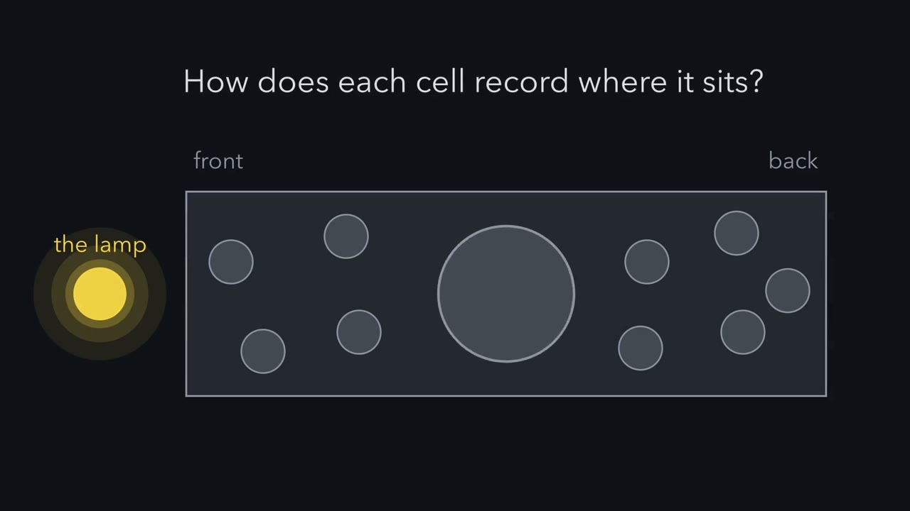
Somatosensory cortex, the touch area, sits in the middle. Inside it lies the barrel field, the whisker map. Auditory cortex sits on the lateral side, toward the back. Visual cortex sits at the back. We will call motor and somatosensory the front-and-middle areas. Visual is the back area. The cortex grows from a smooth sheet of progenitors, cells that divide to make neurons. The sheet that will hold the whisker map and the visual map looks the same from front to back. Making the areas from it is called arealization. Signalling centres at the edges of the sheet are active by about embryonic day 9.5. They keep signalling as the first neurons are made.
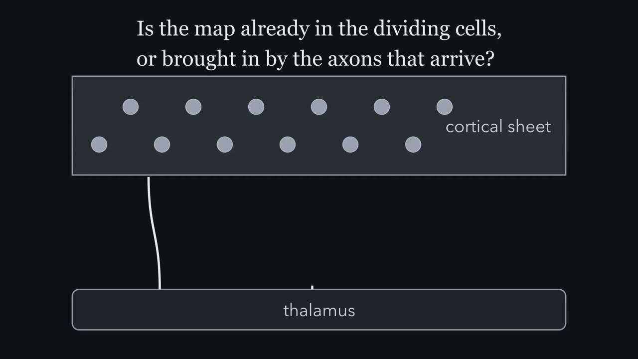
Progenitors make the cortex's neurons from about embryonic day 11 to 17. The thalamus is the relay station for signals from the eyes, ears and skin. Which comes first: the neurons of the cortex, or the thalamic axons that reach them?
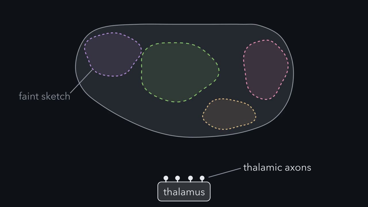
The first neurons are born first. Thalamic axons reach the developing cortex around embryonic day 14 to 15, and wait in the layer just under it. More neurons are still being made then. The axons grow into the cortex itself later, around birth. Barrels can be seen in the first postnatal week, around postnatal day 4. So the sheet is already patterned and making neurons before the thalamic axons arrive. Where do the borders come from, in a sheet that shows none? What tells a progenitor where it sits, and can that be changed so a border moves? And what does input from the thalamus add, if the sheet has already been marked? The takeaway: the areas are real, and the embryonic sheet shows no borders for them.
The next chapters ask what draws them, and when.
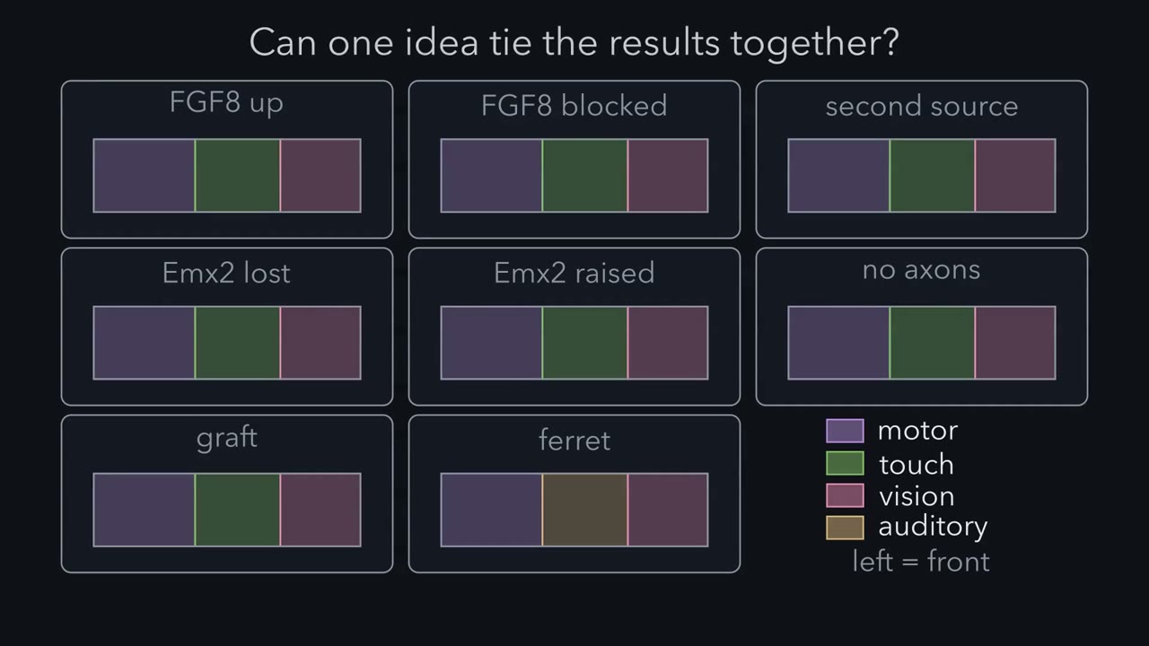
Add one secreted protein to the back of a mouse embryo's cortex and a second, partial whisker map can appear there.
Continue in the lesson: Mapping the Cortex: Areas and Arealization →
3:02 · Free, with captions and a transcript. The lesson has the full text, the questions and the sources.
More films from this lesson
How this film was made. Narration: an AI narrator voice (Kokoro, af_heart) reading narration written from this lesson's text. Animation: AI-generated in code (Manim) by Claude, a schematic with no drawn molecular structures. Captions come from the narration script. See something wrong? Report it.