Cell Signaling · Skim version
TGF-β and Smads
Read the full lesson → Skim the whole course →
The guiding idea
Why does the protein that tells healthy cells to stop dividing end up helping late tumours?
TGF-β sends its message through a short relay.
-
The idea: the ligand only introduces two kinases
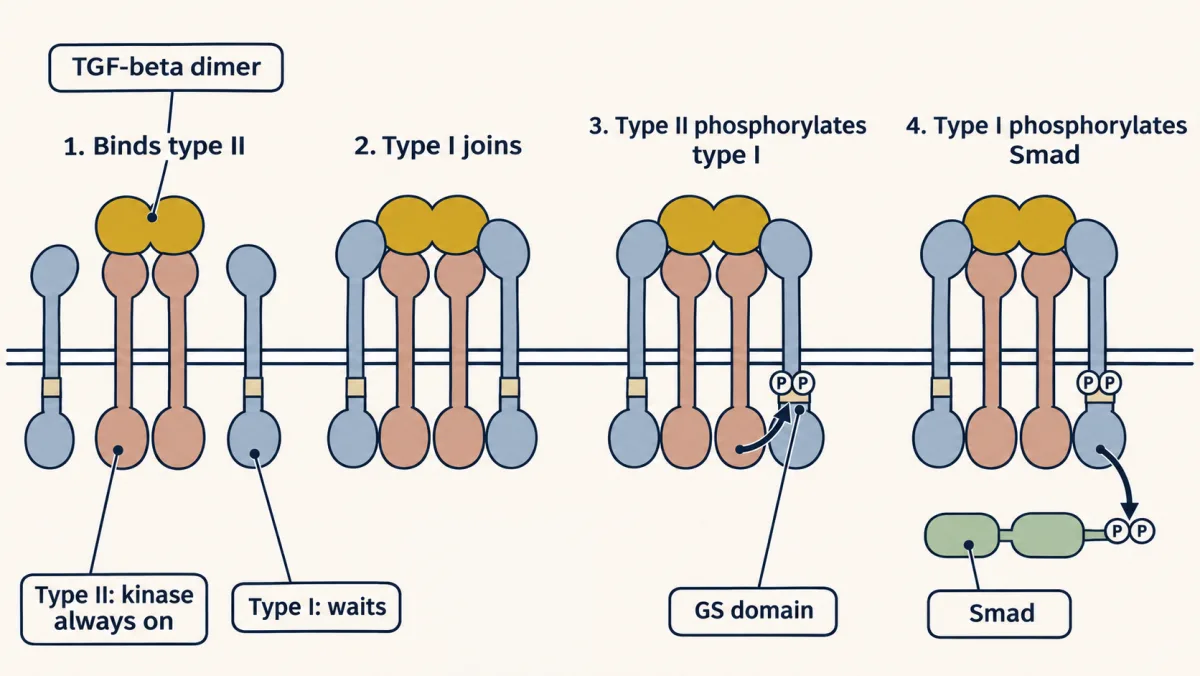
The ligand binds two type II receptors first, then brings in two type I receptors. The always-active type II kinase phosphorylates the GS domain of type I, and the now-active type I kinase phosphorylates a Smad: the ligand's only job is to hold the match to the fuse. AI-generated illustration -
The messenger is the transcription factor
-
Same signal, different answers
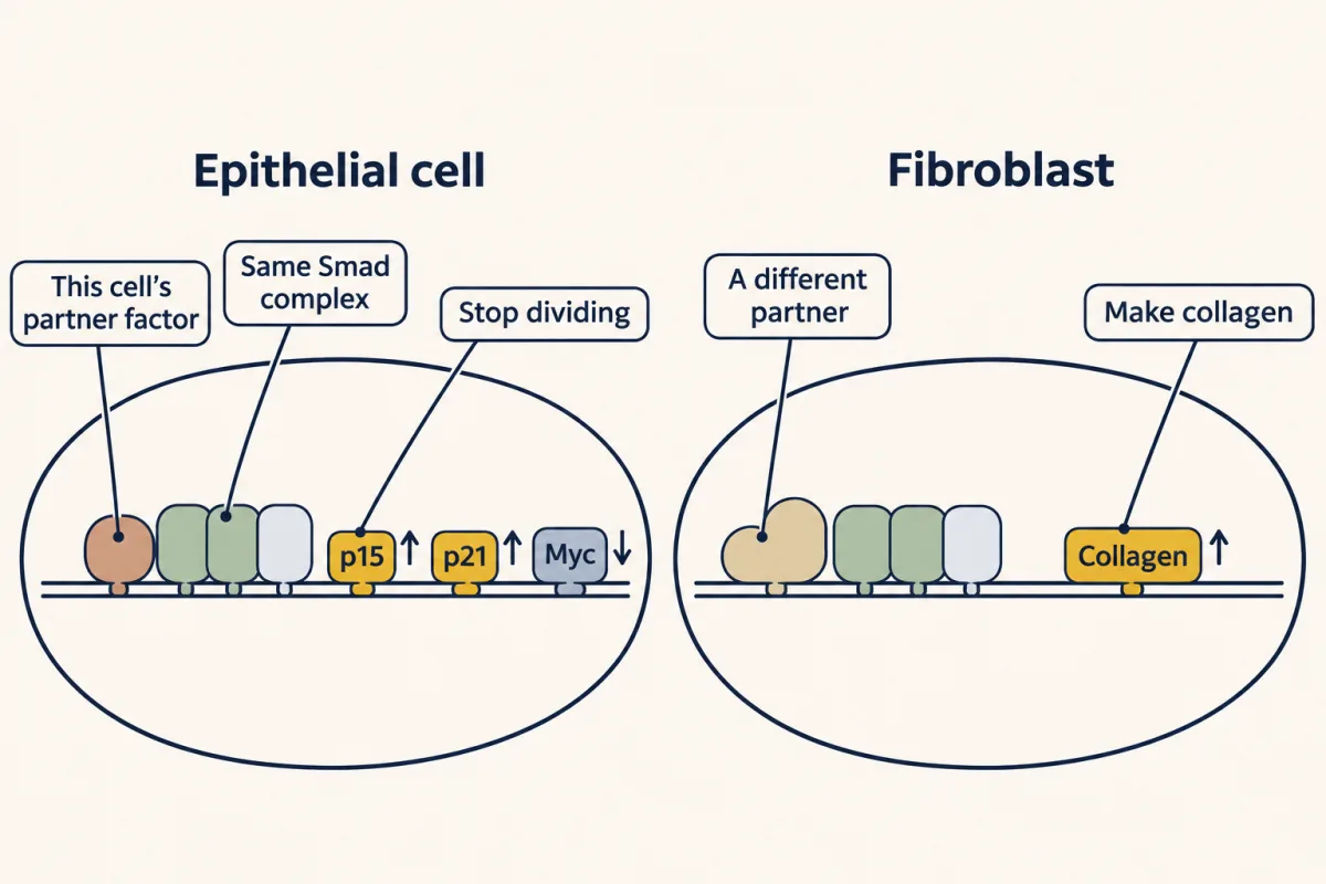
A Smad complex reads sequences too short to pick out a gene on its own; it binds firmly only beside partner transcription factors that each cell type makes for itself. The complex carries one message, that TGF-beta is here, and the partners decide what it means: stop dividing in many epithelial cells, make collagen in a fibroblast. AI-generated illustration -
Three brakes, and what breaks
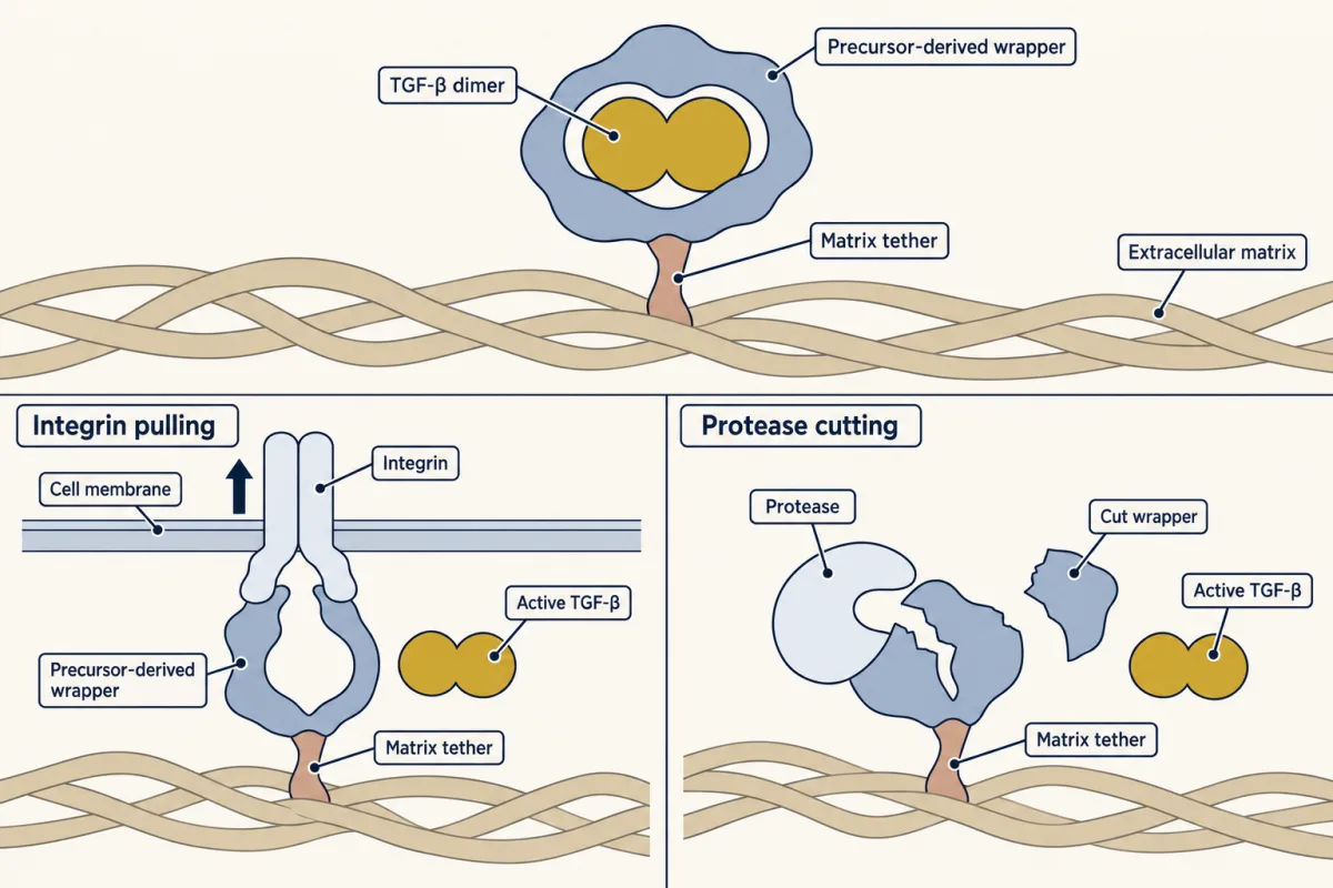
TGF-β is secreted in a latent complex: the cleaved front portion of its precursor remains bound and blocks receptor access. This wrapper is usually tethered to the extracellular matrix. Local activation can occur when cell integrins pull on the wrapper or a protease cuts it, releasing active TGF-β from the wrapper. AI-generated illustration -
Going deeper
Where this lesson fits
Before thisReview first
- Receptor Tyrosine KinasesThis course · Lesson 18
You need the receptor kinase idea from there to see how this serine/threonine receptor pair differs.
- Cytokine Receptors and JAK–STATThis course · Lesson 22
The STAT shortcut, where the messenger is itself a transcription factor, is compared directly with Smads here.
- Ubiquitin and Regulated Proteolysis as SignalsThis course · Lesson 8
Smurfs tag the receptor and Smads for destruction, using the ubiquitin tagging described there.
After thisStudy next
- Wnt: β-Catenin and Planar PolarityThis course · Lesson 24
It takes up another signal whose message ends in gene control and that cancer can corrupt.
- Oncogenes: When the Switch SticksThis course · Lesson 41
It takes further the idea that a broken signalling part, like Smad4 loss, drives cancer.
- The Shape of a Signal: Size, Duration, Frequency and PlaceThis course · Lesson 35
It explains how one cell reads a signal's size and duration, extending why one signal gives different answers.
Elsewhere on the siteFilms, simulations and method films in other courses
- One Rise, One FallFilm · Cell Biology
The cell-cycle brake that TGF-β sets in healthy epithelial cells acts on the cyclin-CDK machinery shown there.
- Cre-lox and binary expression systemsMethod film
Deleting a pathway component only in chosen cells is how its early and late roles in tumours are separated.
Suggested by an AI model from the lessons themselves, then checked. How we use AI