Sensory Systems · Skim version
Parallel Channels Out of the Eye
Read the full lesson → Skim the whole course →
The guiding idea
A hawk's shadow slides across a meadow. What does a mouse's optic nerve tell its brain about it? Not a picture: more than 40 kinds of ganglion cell each report the scene through their own filter.
In the mouse, the optic nerve carries more than 40 overlapping, differently filtered maps of the scene: one ganglion cell type, one mosaic, one map.
From the film: Many Maps, Not One Picture
- The optic nerve carries many overlapping, differently filtered maps of the scene, not one picture.
- In the mouse, each of more than 40 ganglion cell types reports the scene through its own filter, built by retinal circuits before any spike leaves the eye.
- One type, one mosaic, one map: the cells of a type usually keep their distance from one another.
From the film: The Cell That Ignores the World Moving
- A W3 cell fires for a small object moving over a still background and falls nearly silent when the whole scene moves.
- Part of that silencing is presynaptic and part is postsynaptic, and the VG3 amacrine cell supplies much of the excitation.
- The JAM-B cell is a second channel: in isolated retina, its lopsided cells preferred movement from the cell body toward the dendrites.
- Calling W3 cells alarm neurons for overhead predators is a hypothesis, because the experiments used isolated retina.
From the film: One Type or Two? Four Ways to Count
- A real type usually spaces its cells out in its own mosaic, so merging two types usually makes spacing patchy and coverage higher.
- A mosaic is strong support for a type, not a strict rule, because some real types are only weakly regular.
- Imaging, RNA sequencing, spike recording and electron microscopy gave different counts because each reflects a method, a preparation and a resolution, not rival answers.
- The macaque fovea needs its own census, since midget clusters made up about 85% of sampled foveal ganglion cells.
-
One cell that ignores the world moving
-
Testing a type: agreement and a mosaic
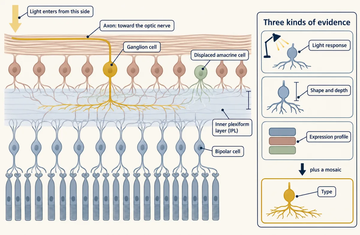
A ganglion cell type is supported by agreement among light response, dendritic shape and depth in the inner plexiform layer, and gene-expression profile. A regular mosaic of cell bodies provides additional support, but regularity varies among types and is not a strict requirement. Dendrites of one type can overlap substantially. Ganglion cell axons join the optic nerve. AI-generated illustration -
Four ways to count
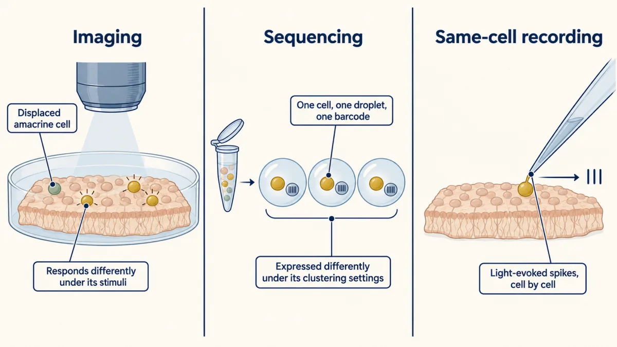
Three of the four mouse retinal censuses, which measured different properties: calcium imaging distinguished at least 32 functional ganglion cell types; RNA sequencing yielded 46 types after a knowledge-guided split of one cluster containing two known direction-selective types; and cell-attached spike recording distinguished 42 functional types, with RNA or morphology recovered from subsets of the same recorded cells. The counts reflect different methods, preparations and resolutions, rather than rival answers to one question. Displaced amacrine cells in the imaged ganglion cell layer were separated from the ganglion cell groups. AI-generated illustration -
Mouse is not macaque: the primate fovea
-
Going deeper
Where this lesson fits
Before thisReview first
- Sensory Neurons by Their Genes: the Atlas EraThis course · Lesson 3
It shows how gene-expression clusters become real neuron types, the same question asked here for ganglion cells.
- Five Ways to Turn a Stimulus into a CurrentThis course · Lesson 1
You need to know how photoreceptors turn light into a current before asking what ganglion cell filters do with that signal.
After thisStudy next
- Computing Motion in the RetinaThis course · Lesson 19
It shows how retinal circuits build the direction-preferring channel previewed here with JAM-B and the direction-selective types.
- Melanopsin Cells: Light for the Clock and the PupilThis course · Lesson 20
Melanopsin cells are a sparse ganglion cell type whose clock and pupil job shows abundance is not importance.
- Calcium Feedback Sets Photoreceptor Gain, and When Photoreceptors DieThis course · Lesson 21
It asks what happens to these ganglion cell channels when photoreceptor input changes or fails.
Suggested by an AI model from the lessons themselves, then checked. How we use AI