Cell Biology · Skim version
Protein Quality Control and Organelle Turnover
Read the full lesson → Skim the whole course →
The guiding idea
When material piles up in a cell, is more arriving or less leaving, and how can you tell?
A crowded quality-control pathway can mean faster arrival or slower clearance.
From the film: Follow the Flow, Not the Pile
- For a defined cellular pool, a larger pool can mean faster arrival or slower clearance.
- In the ER, many failed proteins are returned to the cytosol, tagged with ubiquitin, and destroyed by the proteasome.
- In macroautophagy, autophagosomes carry bulk cytoplasm and whole organelles to lysosomes.
- During starvation, autophagy recycles cell material into reusable building blocks.
- Under a complete lysosomal block that leaves carrier formation unchanged, the extra LC3-II estimates lysosomal turnover.
From the film: When the ER Falls Behind
- Many secreted and membrane protein chains enter the ER and fold with chaperones such as BiP; folded clients can leave for the Golgi.
- A persistently misfolded client can enter ERAD: it is moved toward the cytosol, ubiquitinated, extracted and destroyed by the proteasome.
- When unfolded proteins arrive faster than the ER can clear them, the unfolded protein response slows arrival and raises capacity.
- Mammals have three UPR branches, IRE1, PERK and ATF6; budding yeast relies on one ER-stress sensor, Ire1.
- If ER stress is not relieved, the response can also drive cell death.
-
Physical constraints on the two exits
-
Read a flow, not a still picture
-
The ER: retry, remove, raise capacity
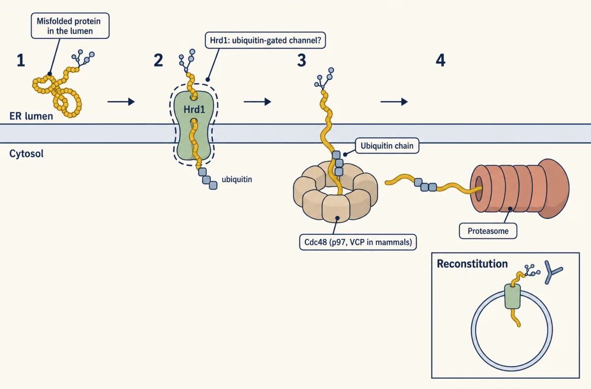
As a failed ER client returns toward the cytosol, it is ubiquitinated, pulled out by Cdc48 and destroyed; the dashed Hrd1 passage is only proposed. AI-generated illustration 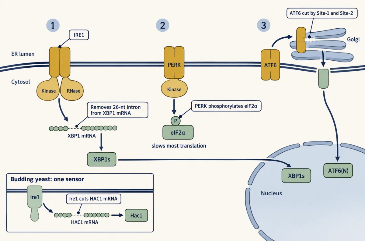
Yeast relies on one ER stress sensor, Ire1, while mammals spread the alarm across three overlapping branches. AI-generated illustration -
Selective autophagy: choosing a mitochondrion
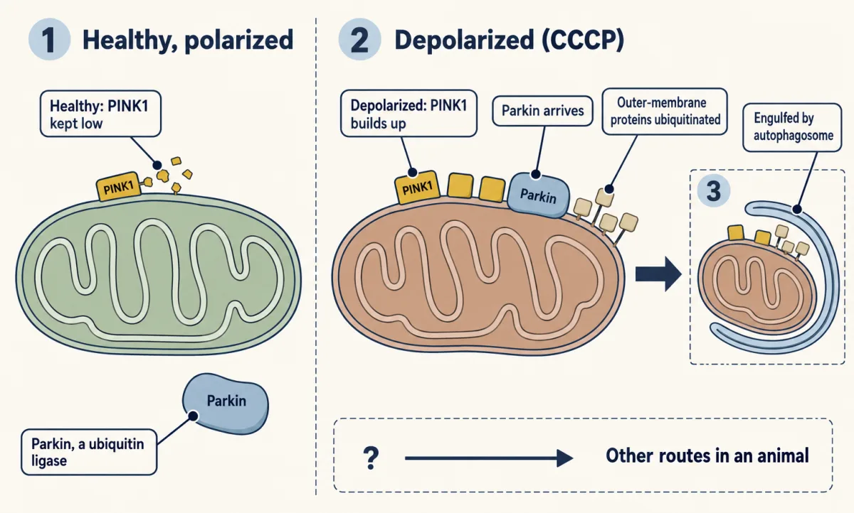
In cultured cells, a depolarized mitochondrion is chosen for autophagy once PINK1 builds up, Parkin arrives and ubiquitin coats its surface. AI-generated illustration -
Going deeper
Where this lesson fits
Before thisReview first
- Protein Sorting into CompartmentsThis course · Lesson 7
You need to know how proteins enter the ER and fold there before asking what happens to failed ones.
- Organelles as Distinct EnvironmentsThis course · Lesson 6
The lysosome's acidic lumen, held by the V-ATPase, is what lets it digest cargo.
After thisStudy next
- Cell Death and ClearanceThis course · Lesson 21
Unrelieved ER stress can push a cell toward death, and this lesson shows how that pathway is carried out.
- Endocytosis, Recycling and DegradationThis course · Lesson 13
Endocytosed material also ends in the lysosome, the same degradation exit autophagy uses.
- Cell Identity, Cell State and DiseaseThis course · Lesson 25
Deciding whether a mutant changes folding, disposal or signalling feeds into interpreting cell state and disease.
Elsewhere on the siteFilms, simulations and method films in other courses
- Choose, Recycle, SplitFilm · Biochemistry
It shows ubiquitin tagging and proteasome degradation, the exit used by failed ER proteins.
Suggested by an AI model from the lessons themselves, then checked. How we use AI