Cell Signaling · Skim version
Nuclear Receptors: Receptors That Are Transcription Factors
Read the full lesson → Skim the whole course →
The guiding idea
What does a cell do with a hormone that goes straight through its membrane?
Steroid and thyroid hormones act through receptors inside the cell that are themselves transcription factors.
-
The idea: the hormone goes in, and its receptor reads DNA
-
One plan, forty-eight receptors
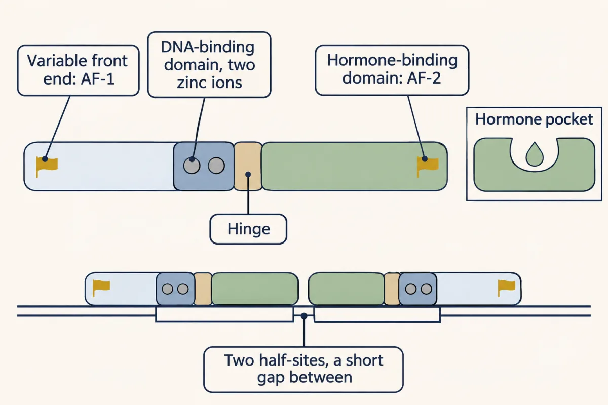
Most of the 48 receptors share one plan: a variable front end with AF-1, a DNA-binding domain folded around two zinc ions, a hinge, and a hormone-binding domain that holds the hormone, pairs two receptors and carries AF-2. Most work in pairs, so their response element has two six-base halves. AI-generated illustration -
Two ways to wait for a hormone
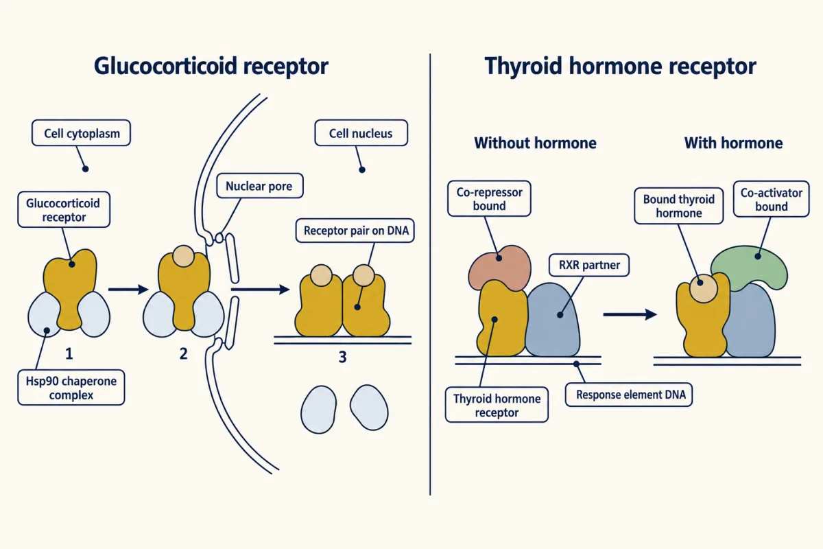
Two examples of how nuclear receptors wait for hormone. The glucocorticoid receptor waits mostly in the cytoplasm with Hsp90 chaperones. Hormone binding rearranges the complex; the receptor enters the nucleus and sheds its chaperones, then pairs and binds DNA. The thyroid hormone receptor waits on DNA with RXR and a co-repressor; hormone binding replaces the co-repressor with a co-activator. This co-repressor model is best documented for thyroid hormone and retinoic acid receptors, rather than every RXR partner. These diagrams show the usual hormone-dependent routes. AI-generated illustration -
What the hormone changes: one helix closes
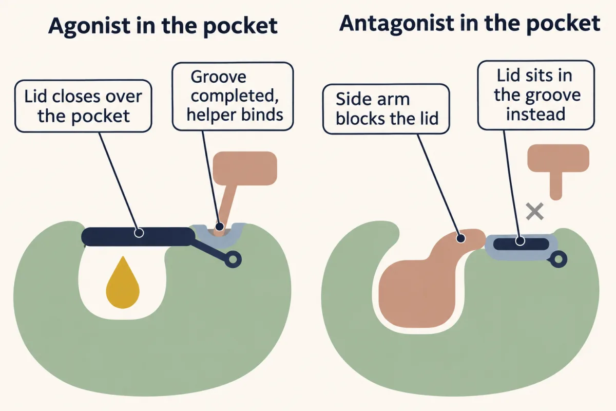
The main moving part is the last helix of the hormone-binding domain. With an agonist it folds across the pocket like a lid and completes the groove a co-activator binds; in the tamoxifen structure a side chain keeps it out and the helix lies in the groove itself, so the groove is taken. AI-generated illustration -
Why it takes hours, and where it meets medicine
Where this lesson fits
Before thisReview first
- A Field Guide to ReceptorsThis course · Lesson 4
It sets out receptor types and why speed follows design, which this lesson uses to place the slow nuclear receptor.
- Binding: Affinity, Occupancy and Dose–ResponseThis course · Lesson 3
You need affinity and occupancy from there to follow how agonists, antagonists and tamoxifen compete for one pocket.
- TGF-β and SmadsThis course · Lesson 23
Smads are the contrasting case where a messenger is the transcription factor, which this lesson refers back to.
After thisStudy next
- Signaling to the Nucleus: CREB, AP-1 and Immediate Early GenesThis course · Lesson 32
It takes the step after receptors reach the nucleus: how signals switch on immediate early genes.
- The Shape of a Signal: Size, Duration, Frequency and PlaceThis course · Lesson 35
It uses the slow, hours-long timing of gene-based signals to ask how cells read a signal's duration.
- Drugs Against a PartThis course · Lesson 42
It extends drug action on one receptor part, as with tamoxifen and the PPAR-γ drugs here.
Suggested by an AI model from the lessons themselves, then checked. How we use AI