Sensory Systems · Skim version
The Cochlear Amplifier and Otoacoustic Emissions
Read the full lesson → Skim the whole course →
The guiding idea
A microphone sealed in a healthy ear canal can record sound that your own cochlea made. What does that sound tell us about the amplifier, and what can it not tell us?
In a living mammalian cochlea, outer hair cells add a gain that is large for soft sounds, small for loud ones and gone after death.
From the film: A Gain That Death Erases
- The cochlear amplifier is the gain of the living cochlea over the dead one, and in mammals it depends on outer hair cells.
- That gain is large for soft sounds, small for loud ones and gone after death.
- Sensitivity, sharp tuning, compressive growth of motion and the returning sound are four signatures of that one gain.
From the film: From a Bent Bundle to a Pushing Cell
- The outer hair cell turns sound into force through a chain: bundle bending, transduction current, voltage, prestin shape change, and a push on the organ of Corti.
- Nonlinear capacitance is the electrical fingerprint of prestin's charge movement, and it does not measure the force or power a hair cell produces.
- In a prestin knock-in mouse amplification was lost while transduction and cell mechanics were normal, so cell-body motility is necessary; how the work is split with the bundle in mammals is not established.
From the film: The Sound That Comes Back
- A fast nonlinearity in a living ear makes new tones, such as 2f1 minus f2, that a microphone in the ear canal can record.
- At screening levels the distortion product fails when outer hair cells fail, which is what ties it to the amplifier.
- A passed emission screen shows outer hair cells can make an emission, not that hearing is intact, because auditory neuropathy can present with a normal emission.
-
A gain that death erases
-
The engine: from a bent bundle to a pushing cell
-
Reading the motor’s fingerprint: nonlinear capacitance
-
What the structures can and cannot show
-
The sound that comes back
-
A second engine? The frog hair bundle
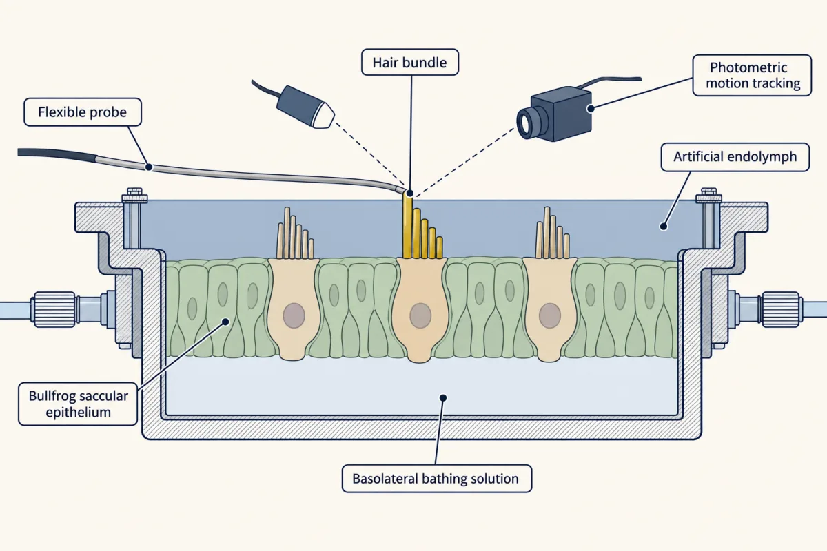
The Martin and Hudspeth rig: excised bullfrog saccular epithelium in a two-compartment chamber, a flexible probe on one hair bundle, and photometry tracking its motion. The inset is the energy comparison that showed the bundle adds energy. AI-generated illustration -
Where along the cochlea is the gain gathered?
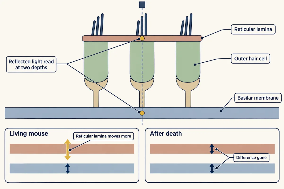
Optical measurements read motion at the reticular lamina and the basilar membrane together. The insets follow Ren and colleagues at one basal site in living and dead mice. AI-generated illustration -
Going deeper
Where this lesson fits
Before thisReview first
- Five Ways to Turn a Stimulus into a CurrentThis course · Lesson 1
You need the hair-cell current and how a bent bundle opens channels from there, since the amplifier chain starts at bundle bending.
- The Hair Cell's Transduction ChannelThis course · Lesson 22
The channel's saturating current is the fast nonlinearity that makes the distortion product, and its current drives prestin.
After thisStudy next
- Hidden Hearing Loss, Gene Therapy and RegenerationThis course · Lesson 24
It takes the vulnerable outer hair cell gain further, to hidden loss after noise and to repair.
- Balance: Two Codes for Every Head MovementThis course · Lesson 25
It uses hair-cell transduction in balance organs, where no outer hair cell amplifier is involved.
Elsewhere on the siteFilms, simulations and method films in other courses
- The permeability cliffSimulation · Biochemistry
Prestin's charge-moving two-state behaviour rests on how a membrane's electric field acts on proteins in the bilayer.
Suggested by an AI model from the lessons themselves, then checked. How we use AI