Biochemistry · Skim version
Amino Acids: How One Side Chain Decides Everything
Read the full lesson → Skim the whole course →
The guiding idea
The R group sets what makes each amino acid different — its charge, its pKa, and the Glu→Val swap behind sickle cell.
Twenty amino acids share one backbone, so what makes them different — and why can a single swap among them turn a healthy protein into a disease?
From the film
- Each ionizable group is a proton switch: below its pKa it mostly holds its proton, above it mostly lets go.
- Holding a proton makes a group one charge more positive: acid-type switches go from 0 to −1, base-type from +1 to 0.
- An amino acid's net charge is the sum of its switches; at pH 7.4, glycine is a net-zero zwitterion.
- The pI is the pH of zero net charge; for glycine it is the average of its two pKa values, about 5.97.
-
One backbone, twenty side chains
-
Proton switches: the one rule for charge
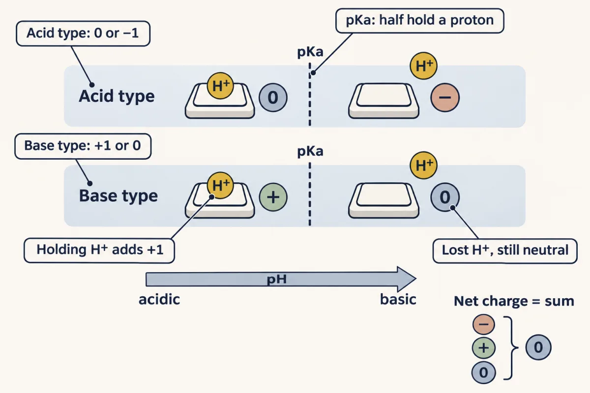
Every ionizable group is a proton switch. Below its pKa it mostly holds H+; above it, it mostly lets go. Holding the proton always makes the group one charge more positive, so an acid-type group goes from 0 to −1 and a base-type group from +1 to 0. An amino acid's net charge is the sum of its switches. AI-generated illustration -
Four side-chain families, read from the switches
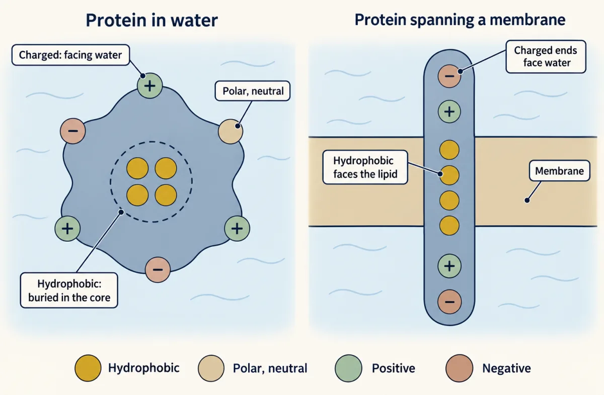
Side-chain properties help predict residue location. In water-soluble proteins, hydrophobic side chains tend to be buried, while polar and charged side chains often face water. In membrane-spanning regions, lipid-facing side chains are predominantly hydrophobic. These schematics show representative arrangements. AI-generated illustration -
Reading the curve: titration, pKa and pI
-
Electrophoresis: which way does it move?
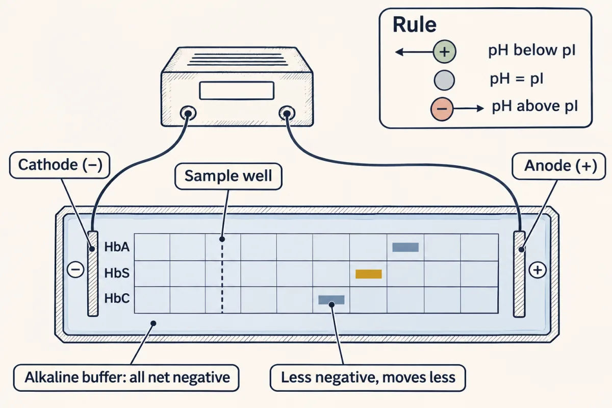
In electrophoresis a molecule moves according to its net charge. Below its pI it is positive and heads for the cathode; above its pI it heads for the anode. In an alkaline buffer every hemoglobin is net negative, but relative to HbA, HbS is two charges more positive per molecule and HbC four, so toward the anode the order is HbA, HbS, HbC. AI-generated illustration -
The payoff: one swap, two properties
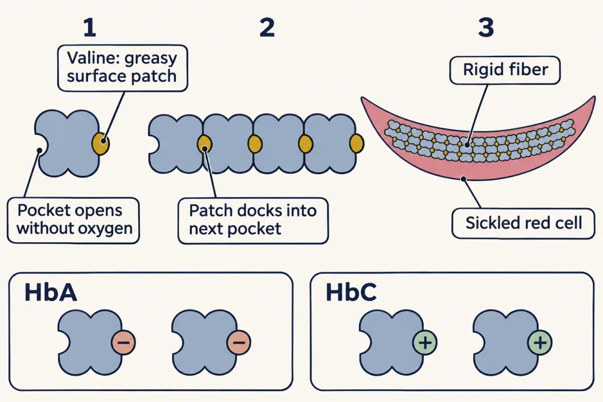
Why cells sickle is a polarity story. Valine puts a hydrophobic patch on the surface of HbS; when hemoglobin releases oxygen, a matching hydrophobic pocket opens, and the patch of one molecule docks into the next, building rigid fibers that deform the red cell. Glutamate (HbA) and lysine (HbC) are both charged and hydrophilic, so neither plants the patch. AI-generated illustration -
Going deeper
Where this lesson fits
Before thisReview first
- Water, Weak Bonds, and the Generation of OrderThis course · Lesson 3
You need its account of water and weak bonds to see why polar side chains face water and greasy ones hide.
After thisStudy next
- Protein Three-Dimensional Structure: The Sequence Sets the ShapeThis course · Lesson 5
It uses these side-chain properties to explain how a sequence folds, and why one swap can break the shape.
- Protein Binding and Allostery: How Hemoglobin Loads and Delivers OxygenThis course · Lesson 9
It takes the Glu→Val swap and histidine's proton switching further, using hemoglobin's oxygen binding and release.
- Techniques in Protein Biochemistry: Purify It, Then Prove ItThis course · Lesson 6
It uses each protein's charge at a given pH and its pI to separate proteins on columns and gels.
Elsewhere on the siteFilms, simulations and method films in other courses
- Build a big molecule from small blocksSimulation · Biology
Building a big molecule from small blocks shows how amino acids link into chains before side chains matter.
- Take It Apart, Build It AgainFilm · Biology
It shows amino acids as the building blocks that digestion splits and the body rejoins into protein.
- Speed up and slow down an enzymeSimulation · Biology
Enzyme activity changes with pH because charged side chains in the active site gain or lose protons.
Suggested by an AI model from the lessons themselves, then checked. How we use AI