Films · Developmental Neuroscience
Two Routes to a Neuron
Read the transcript of “Two Routes to a Neuron”
Film transcript: Two Routes to a Neuron
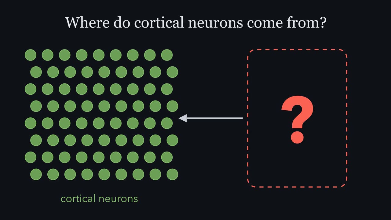
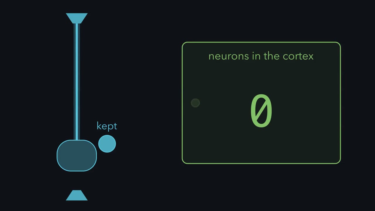
In an asymmetric division, a radial glial cell keeps one daughter. What happens to the other one? It can take one of two routes. In direct neurogenesis, the radial glial cell divides into another radial glial cell and a neuron. In indirect neurogenesis, the second daughter is an intermediate progenitor. It lets go of the ventricle and moves into the subventricular zone. There it divides once more, in rodent cortex usually into two neurons. Filming rat slices, Noctor and colleagues tracked both routes. Most radial glial divisions were asymmetric. About nine in ten intermediate progenitor divisions gave two neurons. But only 19 divisions away from the ventricle were seen, so read it as rat.
In mouse, intermediate progenitors may make most cortical projection neurons. Transcription factors, proteins that switch genes on or off, mark the steps. Pax6 marks radial glia. Tbr2 marks intermediate progenitors. Tbr1 marks young neurons. They appear in that order, as cells move outward. But are the markers only labels? In 2008, two groups removed Tbr2 from the developing mouse brain. Count mitoses at the ventricle, and away from it. If Tbr2 is a step in the indirect route, which count should fall?
Division at the ventricle went on. Division away from it fell. And fewer cortical neurons were made. The position of mitosis, read with the markers, pointed to the lost progenitor. In those mice, Tbr2 was needed for the indirect route. Primates have a third progenitor in far greater numbers. Outer radial glia divide far from the ventricle. Mouse has a few. Humans have far more. Now, what does a leaving daughter give up? A niche is the local setting that keeps a stem cell a stem cell. The apical foot faces the cerebrospinal fluid. In mouse, that fluid carries growth factors, among them IGF2, that support proliferation. The basal foot rests on the basement membrane under the pia.
Neighbouring cells, through Notch, are part of the setting too. In an asymmetric division, the daughter that leaves lets go of the apical surface. It gives up the fluid side of the niche, and its growth signals. The daughter that stays keeps it. Losing apical contact goes with leaving, but it is not the same thing. An intermediate progenitor has let go, and still divides once more. So a spent daughter makes one neuron directly, or usually two through an intermediate progenitor. Follow its markers and divisions to tell which.
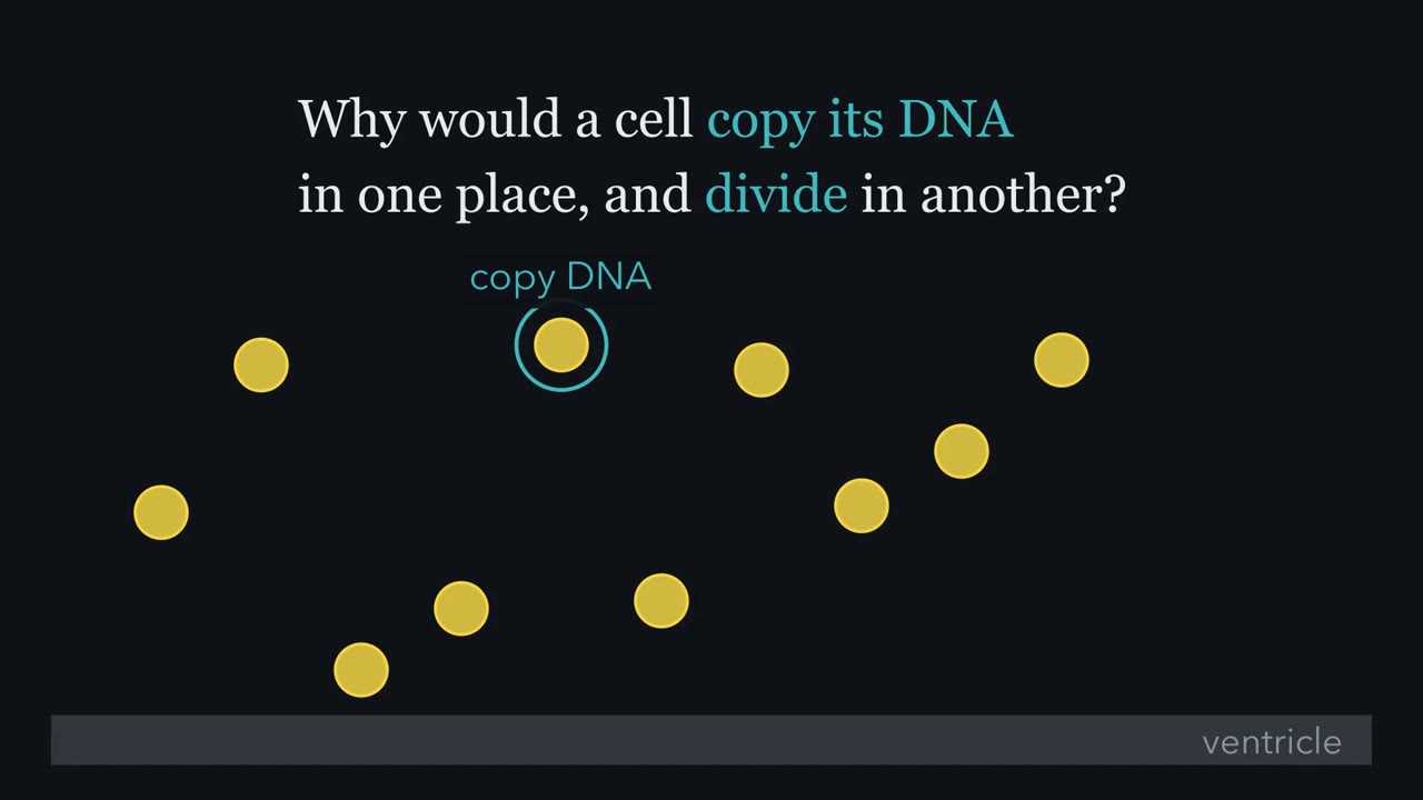
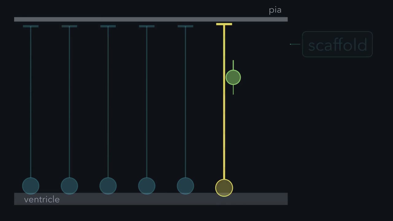
In rodents, radial glia, long seen as mere scaffolding, make most cortical neurons. Why does such a progenitor copy its DNA in one place and divide in another?
Continue in the lesson: The Ventricular Zone: Radial Glia as Stem Cells →
3:10 · Free, with captions and a transcript. The lesson has the full text, the questions and the sources.
More films from this lesson
How this film was made. Narration: an AI narrator voice (Kokoro, af_heart) reading narration written from this lesson's text. Animation: AI-generated in code (Manim) by Claude, a schematic with no drawn molecular structures. Captions come from the narration script. See something wrong? Report it.