Films · Developmental Neuroscience
Scaffold or Source?
Read the transcript of “Scaffold or Source?”
Film transcript: Scaffold or Source?
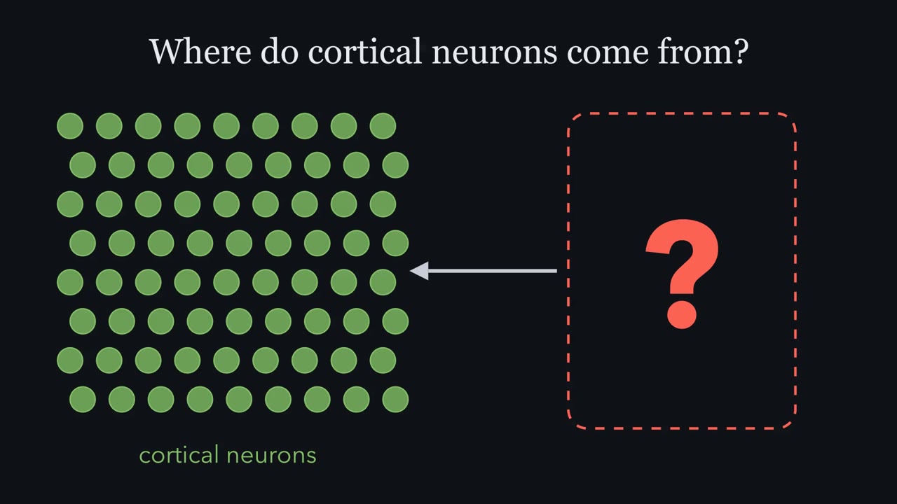
Radial glial fibres guide young neurons outward. Are those same cells also the source of the neurons? In 1972, Pasko Rakic studied fetal rhesus monkey cortex. He found young neurons pressed against radial fibres. Radial glia were seen mainly as scaffolds. Neurons, it was thought, came from some other cell in the ventricular zone. Around the year 2000, three lines of evidence in rodents, using four methods, changed the picture. Each asked the question a different way. For each one, ask: if radial glia were only a scaffold, what would we see? First, sorting, in mouse. The Götz group used mice that make GFP from a human GFAP promoter. Rodent radial glia make little GFAP protein.
But that human promoter fragment is active in them. The green cells were sorted into a dish. If they were only scaffolds, you might expect few or no neurons. Did they make any? In culture, they made neurons. But a dish shows potential, not fate. Second, fate mapping in the living mouse. An enzyme called Cre recombinase was made from a GFAP promoter. Cre cuts out a stop sequence in front of a reporter gene. The cut is a change in the DNA. So it is copied into every descendant, even after the promoter switches off in a neuron. If radial glia were only scaffolds, cortical neurons would stay dark. What did the fate map show?
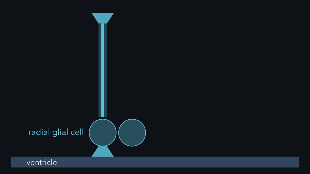
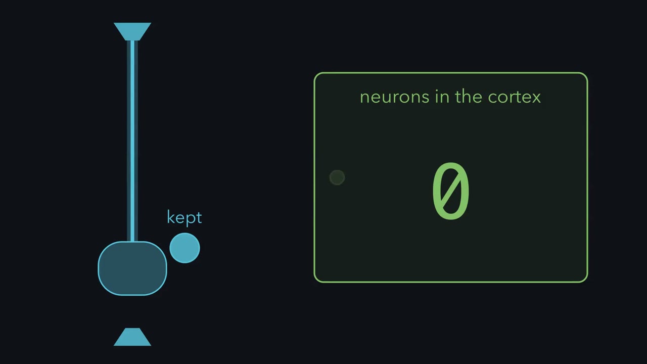
Nearly all cortical projection neurons carried the reporter. Cortical interneurons, local inhibitory cells, did not. They are born in the ventral forebrain. Third, filming lineage, in rat. A retrovirus carrying GFP was injected into the ventricles. This retrovirus enters only dividing cells, and these cells divide right at the ventricle. Every descendant of a marked cell inherits the mark. A green radial glial cell divides at the ventricle. It keeps its fibre. Its daughter becomes a neuron and moves out along a parental fibre. In mouse, Miyata and colleagues saw a newborn neuron inherit the fibre itself. Last, recording, in rat. Precursors were recorded with an electrode, then filled with dye.
The filled cells had the shape of radial glia. Radial glial markers labeled nearly all cells caught in S phase or mitosis. Each method has a blind spot. A dish shows potential. An electrode samples only cells it can reach. A marker is not a family tree. But the blind spots differ. And every result went against a scaffold-only view. The fate map and the films showed neuronal ancestry directly. So in embryonic rodent cortex, the cell that guides migrating neurons is also the cell that makes most of them.
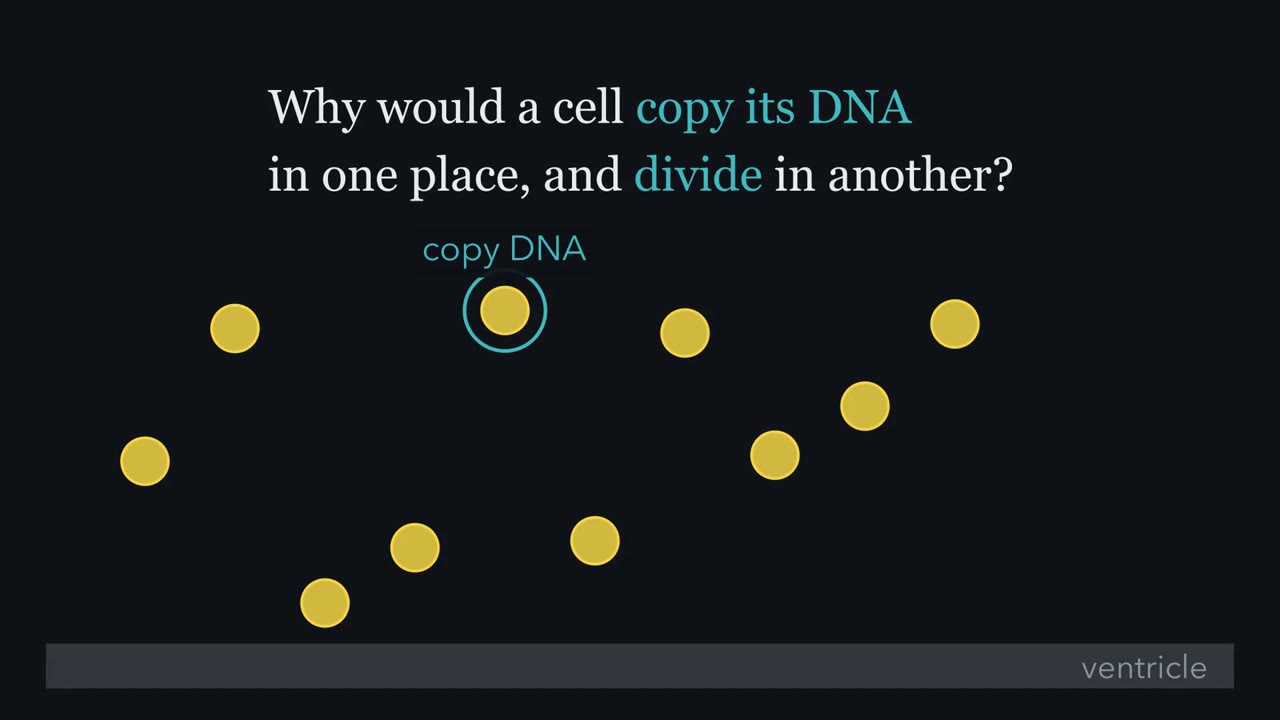
In rodents, radial glia, long seen as mere scaffolding, make most cortical neurons. Why does such a progenitor copy its DNA in one place and divide in another?
Continue in the lesson: The Ventricular Zone: Radial Glia as Stem Cells →
3:26 · Free, with captions and a transcript. The lesson has the full text, the questions and the sources.
More films from this lesson
How this film was made. Narration: an AI narrator voice (Kokoro, af_heart) reading narration written from this lesson's text. Animation: AI-generated in code (Manim) by Claude, a schematic with no drawn molecular structures. Captions come from the narration script. See something wrong? Report it.