Films · Developmental Neuroscience
The Lift: A Nucleus on a Timetable
Read the transcript of “The Lift: A Nucleus on a Timetable”
Film transcript: The Lift: A Nucleus on a Timetable
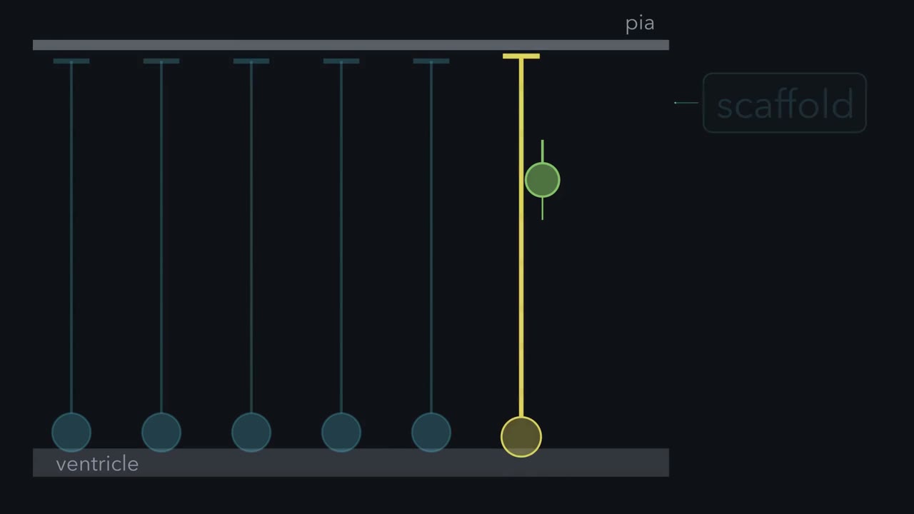
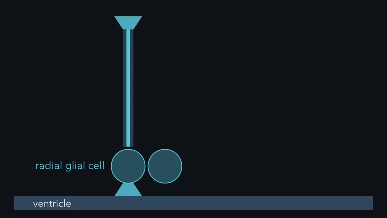
Why would a cell copy its DNA in one place, and divide in another? Nineteenth-century anatomists saw nuclei at many heights. They read the wall as several kinds of cells. In 1935, F. C. Sauer argued for a simpler picture. One kind of long cell, whose nucleus moves inside it. A single layer of cells with nuclei at many heights is called a pseudostratified epithelium. Picture a lift, with the ventricle at the bottom. The shaft is the cell. The car is the nucleus. The shaft runs all the way up to the pia. But the car only serves the lowest floors, the ventricular zone. In G1, the car rides up. It stops near the top of the ventricular zone for S phase, when DNA is copied.
In G2, it comes back down to the ground floor, the apical surface. The cell divides there. Each cell's car runs on its own timetable. That is why nuclei sit at every height. This movement is called interkinetic nuclear migration. Movement between divisions. A pulse label made Sauer's cycle a timetable. Tritiated thymidine, a radioactive DNA building block, is taken up only in S phase. Give a brief pulse, then fix the tissue at intervals. Soon after the pulse, where are the labeled nuclei? And when do labeled mitoses appear?
Soon after the pulse, labeled nuclei sit out in the ventricular zone. No cell in mitosis is labeled. Later, the labeled nuclei come down and divide at the ventricle. In 1959, two groups reported this. Labeled nuclei moved in step with the cell cycle. In eleven-day mouse embryos, no labeled mitosis appeared until an hour after the injection. That lag roughly spans G2, which includes the trip back down. So why divide at the ventricle? The answer is partly settled. The best-supported part. For much of interphase, the centrosome sits at the apical surface, at the base of the primary cilium. A dividing cell builds its spindle from its centrosomes.
On the usual account, the nucleus travels to meet them. Late in G2, the centrosome can also move to meet the nucleus. One proposal is packing. Only a cell in mitosis is round and wide. The rest can stay thin at the surface. A round cell takes about twenty times the room of a thin foot. So on average, each cell needs about a tenth of a dividing cell's room. Staggered timetables let more progenitors share the ventricle. A second proposal is signals. In zebrafish retina, Notch signalling was reported to fall from apical to basal. So how far a nucleus travels could change the signal it meets. Now the payoff. Apical radial glia divide at the surface.
So where a cell divides is a first clue to what kind it is. Most mitoses at the ventricle are apical radial glia, though some intermediate progenitors divide there too. Mitoses away from it are mostly intermediate progenitors, or outer radial glia. To name the cell, add its shape and markers. To learn what its daughters become, you need lineage, the descendants of one cell.
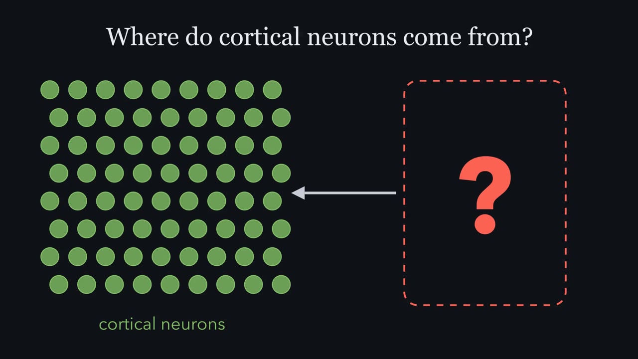
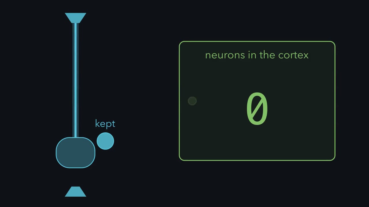
In rodents, radial glia, long seen as mere scaffolding, make most cortical neurons. Why does such a progenitor copy its DNA in one place and divide in another?
Continue in the lesson: The Ventricular Zone: Radial Glia as Stem Cells →
3:49 · Free, with captions and a transcript. The lesson has the full text, the questions and the sources.
More films from this lesson
How this film was made. Narration: an AI narrator voice (Kokoro, af_heart) reading narration written from this lesson's text. Animation: AI-generated in code (Manim) by Claude, a schematic with no drawn molecular structures. Captions come from the narration script. See something wrong? Report it.