More Than a Map: Timing in the Bulb
Read the transcript of “More Than a Map: Timing in the Bulb”
Film transcript: More Than a Map: Timing in the Bulb
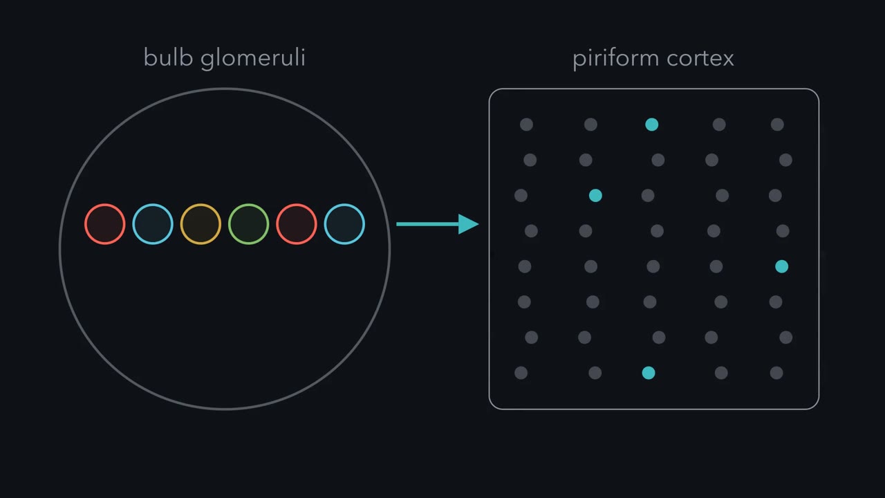
So far the bulb's output was a set of active glomeruli. Is that all the cortex receives? No. The bulb reshapes the pattern with inhibition, and the pattern carries timing. The key contact is a reciprocal dendrodendritic synapse, one dendrite signalling to another and back. A mitral or tufted dendrite releases glutamate onto the interneuron. The interneuron releases GABA back onto the same dendrite. The loop can inhibit the cell that excited it. That is recurrent inhibition. One interneuron touches many output cells, so it can also inhibit neighbours. That is lateral inhibition. Granule cells are not the only source of this inhibition. This resembles the lateral inhibition of sensory coding.
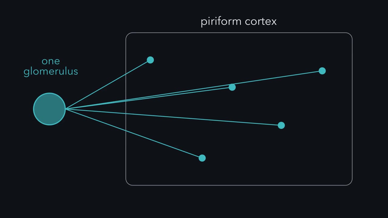
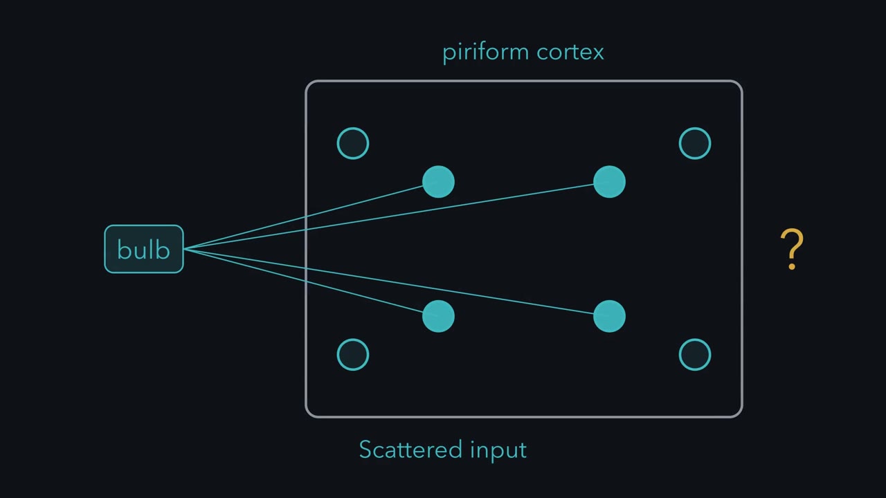
But the bulb is not a retina. Each mitral cell is influenced by a few scattered glomeruli, not a ring of neighbours around it. Now timing. A sniff is a cycle of breathing in and breathing out. A spike's phase is where in that cycle it falls. A review of bulb coding concluded that the relative timing of activity in the bulb conveys odour information. Whether and how downstream circuits use that timing has been debated. Tufted and mitral cells lock to opposite phases of the sniff. They are parallel channels, not copies. Sister cells make a sharp test. Do cells that share one glomerulus send the same message? Dhawale and colleagues recorded in anaesthetized adult mice.
A light-based method found which recorded cells received input from one parent glomerulus. So sisters were defined by their input, not by how they responded to odours. Pause and predict. Sisters share one input. Will their rates match? Will their timing against the breath match?
Counting mean spikes, sister responses were correlated. But against the breathing cycle, an odour shifted the timing of sisters differently. Rate made them look alike. Timing did not. A shared glomerulus is a shared address, not a shared message. Read this within its limits. The mice were anaesthetized, and mitral and tufted cells were pooled. So some timing differences may reflect cell type or state. So the cortex receives a timed map shaped by inhibition. It turns that into a distributed code that its network reshapes and that drifts.
The bulb hands piriform cortex a tidy map of receptor identity, and the cortex scatters it. How can a scattered pattern still carry which odours are alike, and come to mean something?
Continue in the lesson: From Glomeruli to Piriform Cortex →
2:54 · Free, with captions and a transcript. The lesson has the full text, the questions and the sources.
More films from this lesson
How this film was made. Narration: an AI narrator voice (Kokoro, af_heart) reading narration written from this lesson's text. Animation: AI-generated in code (Manim) by Claude, a schematic with no drawn molecular structures. Captions come from the narration script. See something wrong? Report it.