Films · Developmental Neuroscience
The Organizer's Catchers
Read the transcript of “The Organizer's Catchers”
Film transcript: The Organizer's Catchers
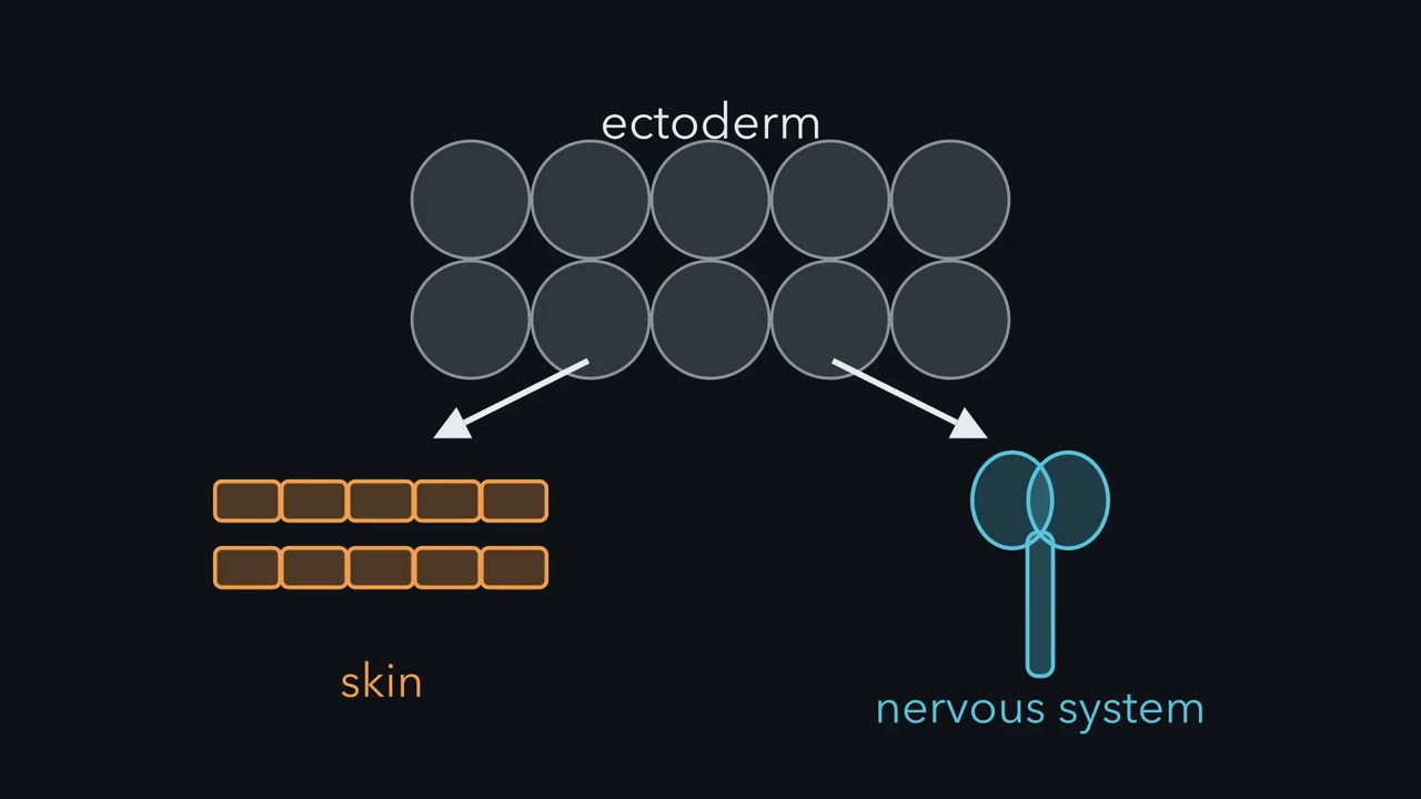
If BMP makes skin, what should the organizer make to turn ectoderm neural? Something that blocks BMP. It makes at least three such proteins. First, where they come from. Chordin and Noggin are made early, at the blastula stage, in dorsal animal cells. So neural induction starts before gastrulation. Then the organizer and dorsal mesoderm turn inward and spread beneath the ectoderm. This is involution.
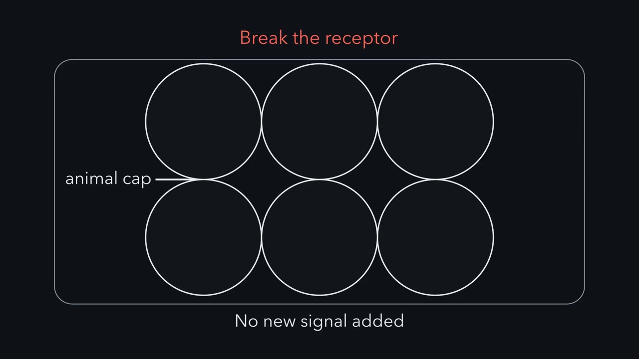
The involuting mesoderm secretes all three. The ectoderm above it becomes the neural plate. Noggin was found in 1992 by William Smith and Richard Harland. It is a secreted protein made in the organizer. In 1993, Teresa Lamb, Harland and colleagues added Noggin protein to intact caps. What should the caps become?
Neural tissue, without dorsal mesoderm. Noggin acted on the ectoderm directly. The markers were all anterior. Forebrain is neural. Cement gland, a mucus gland at the head end, is not. Chordin was cloned in 1994 by Yoshiki Sasai and colleagues, in Eddy De Robertis's laboratory. Both Noggin and Chordin bind BMP4 and keep it off its receptors. Follistatin was known as a protein that binds activin. It also neuralizes caps and binds some BMPs. It works differently. It can stay bound to BMP as BMP docks on its receptor. That complex does not signal. None of the three needs to enter a cell. Each grabs BMP outside the cell. In the crystal structure, Noggin clasps a BMP dimer and covers the faces the receptors would bind.
Chordin has a further twist. In the shuttling model, BMP bound to Chordin travels toward the ventral side. There, Tolloid proteases cut Chordin and release the BMP. Caps show what the catchers can do. Does the embryo need them? In 2005, Khokha and colleagues depleted Chordin, Noggin and Follistatin together from frog embryos. What happens to the neural plate?
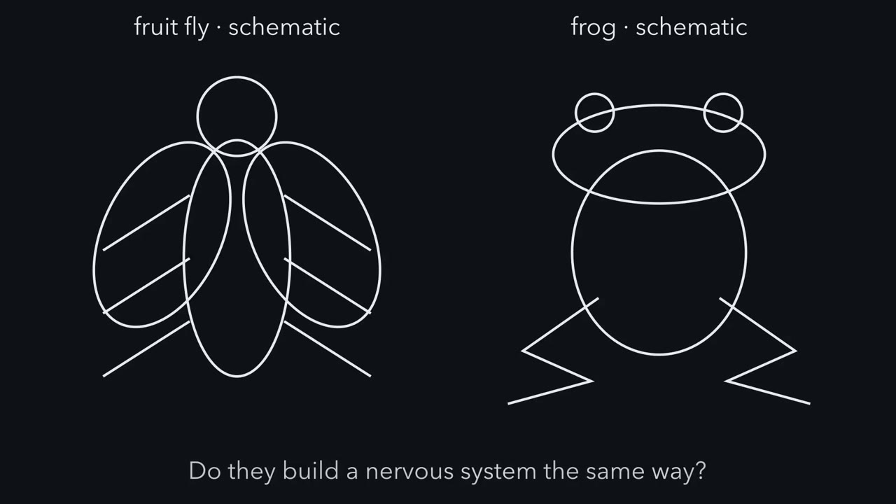
The embryos lost their dorsal structures, the neural plate among them. With all three catchers gone, BMP signaling stays high. The reverse test agreed. Reversade's group removed three BMPs from embryos whose organizer signals were blocked. The embryos formed massive amounts of brain. Early FGF signaling is also needed in the frog, in addition to blocking BMP. It can turn the dial down through MAPK. It also does more.
The takeaway: the frog embryo needs the catchers to keep the dial down. And FGF adds a second input.
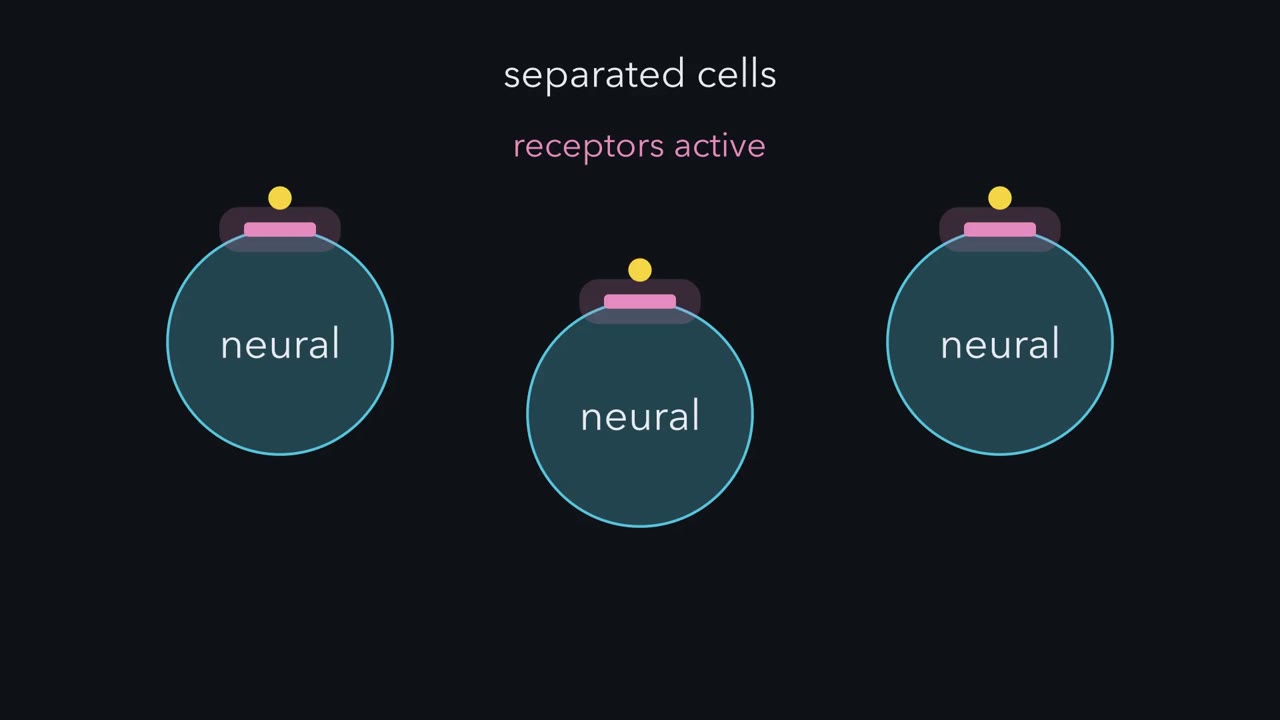
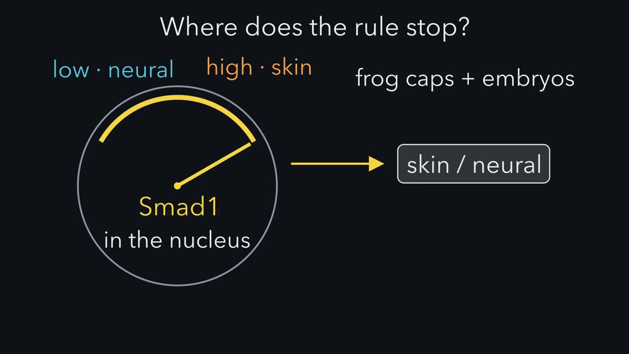
Pull a square of frog skin-to-be apart into single cells, and it becomes neural. Why? Because BMP signaling, which makes skin, falls in those cells. In the embryo, the organizer's secreted proteins make it fall.
Continue in the lesson: Neural Induction: The Default Model →
3:29 · Free, with captions and a transcript. The lesson has the full text, the questions and the sources.
More films from this lesson
How this film was made. Narration: an AI narrator voice (Kokoro, af_heart) reading narration written from this lesson's text. Animation: AI-generated in code (Manim) by Claude, a schematic with no drawn molecular structures. Captions come from the narration script. See something wrong? Report it.