Neuroscience · Skim version
The Retina
Read the full lesson → Skim the whole course →
The guiding idea
A rod in the dark is depolarized and releasing glutamate. A flash of light turns it down. How does quieting a cell let you see?
Light quiets each photoreceptor, but one receptor cannot separate wavelength from intensity, so the retina's circuit compares signals to report lighter or darker, edges and colour.
From the film: One Cell Counts
- Rods and cones are turned down by their stimulus: a photon closes cGMP-gated channels, the cell hyperpolarizes, and its glutamate release falls.
- In dim light a rod counts photons, since each event of about 1 pA in a toad rod is one photoisomerization.
- Once absorbed, every photon has the same effect whatever its wavelength, so one photoreceptor cannot separate wavelength from intensity and the circuit compares.
From the film: One Transmitter, Two Signs
- Glutamate's sign is set by the receptor, not the molecule: OFF bipolar cells use ionotropic receptors and keep the sign, while ON bipolar cells use mGluR6 and invert it.
- Counting the sign inversions from the photoreceptor shows which cells are ON: an even number means the cell responds like light.
- In mudpuppy retina, a glutamate analogue abolished ON responses and spared OFF responses, showing that the two cell types read glutamate with different receptors.
From the film: Centre Minus Surround
- A ganglion cell has a centre and an opposing surround, from bipolar cells and from lateral horizontal and amacrine cells, so it reports contrast more than overall light level.
- In the lesson's linear example, light on both centre and surround gives fewer spikes than the best centre spot, because the surround subtracts only part of the centre.
- Across an edge, in a linear model, ON-centre cells just on the bright side fire most, so the edge stands out across the population.
- Colour comes from comparing cones with overlapping sensitivity curves, and a third cone class breaks ties that two classes would confuse.
-
The layout
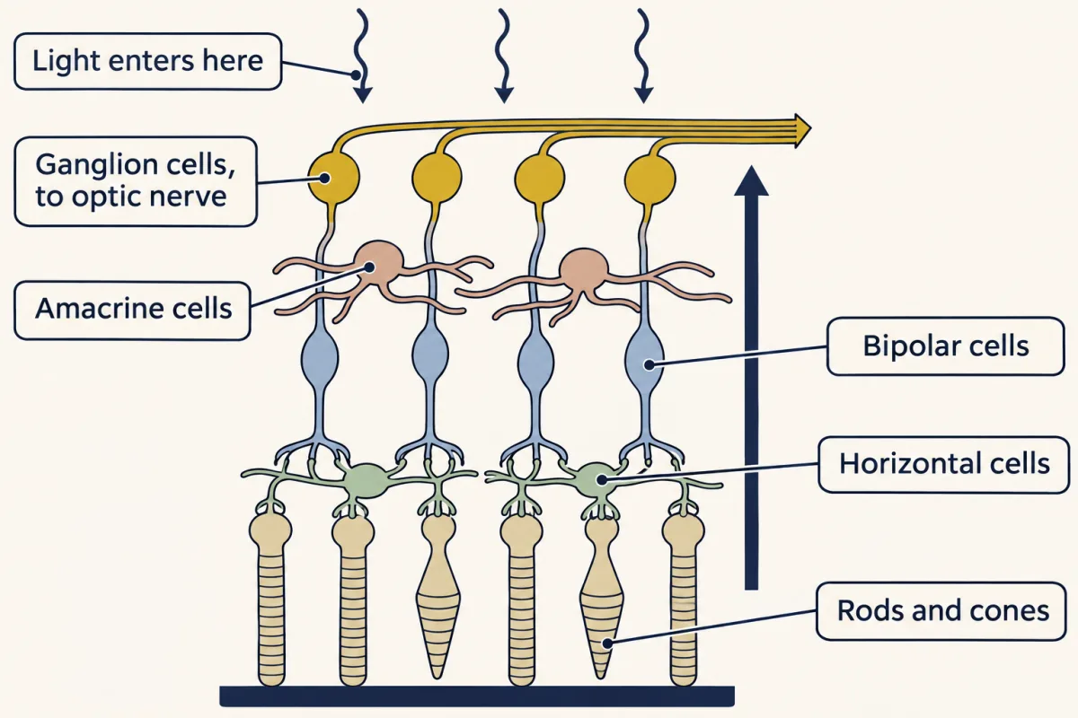
Light crosses the inner retina to reach the rods and cones at the back, and the signal runs back through bipolar cells to the ganglion cells, with horizontal and amacrine cells working sideways. AI-generated illustration -
Light turns the photoreceptor down
-
Counting photons
-
One transmitter, two signs
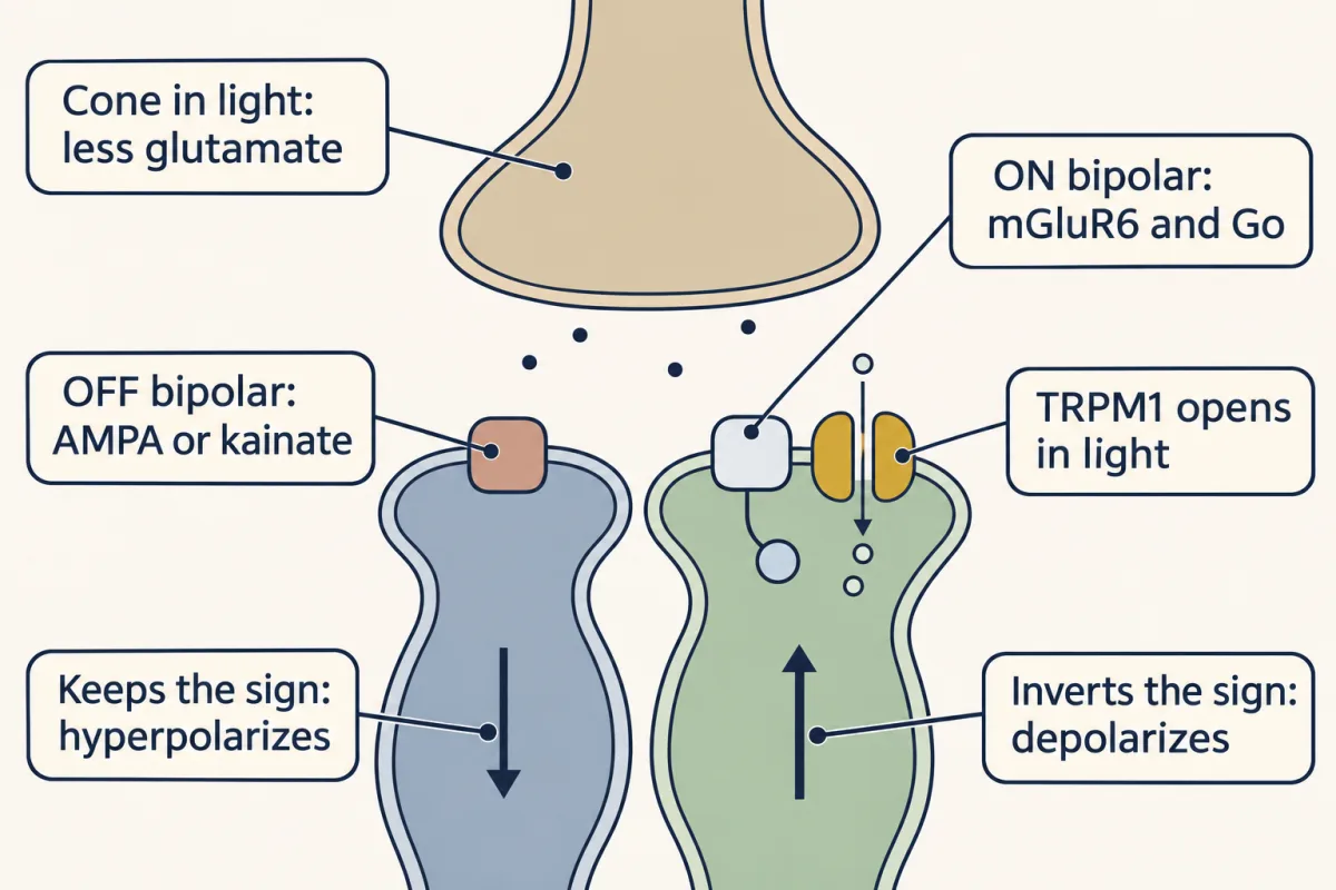
One transmitter, two signs: when light lowers a cone's glutamate release, OFF bipolar cells hyperpolarize, keeping the sign, and ON bipolar cells, whose TRPM1 channels open, depolarize, inverting it. AI-generated illustration -
Centre minus surround
-
Rods borrow the cone circuits
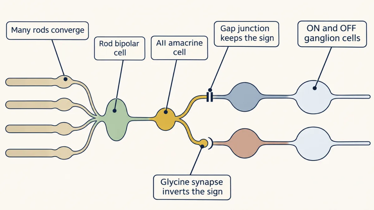
In the primary rod pathway, as mapped in rabbit retina, many rods feed one rod bipolar cell, and the AII amacrine cell passes the signal to the cone ON line by a gap junction and to the OFF line by a sign-inverting glycine synapse. AI-generated illustration -
Colour is a comparison
-
Going deeper
Where this lesson fits
Before thisReview first
- The Resting Membrane and Passive SignalsThis course · Lesson 3
You need the balance point of conductances there to see why closing a cation channel makes the photoreceptor more negative.
- Principles of Sensory CodingThis course · Lesson 9
It introduces receptive fields and graded responses, which this lesson uses to read centre and surround.
- Synaptic Transmission and Quantal ReleaseThis course · Lesson 5
Graded glutamate release and Poisson quantal counting from there are reused for photoreceptor terminals and photon counts.
After thisStudy next
- Visual CortexThis course · Lesson 11
It follows ganglion cell output through the optic chiasm to cortex, where centre-surround fields build orientation selectivity.
Elsewhere on the siteFilms, simulations and method films in other courses
- A Veto From the Null SideFilm · Sensory Systems
It shows how amacrine-cell inhibition builds a ganglion cell's preference for one motion direction.
- A Current That Light Can Use UpFilm · Sensory Systems
It takes the calcium feedback and GCAP gain control of photoreceptors further, with the dark current.
- A Fall Can Carry a SignalFilm · Cell Signaling
It shows the rod cascade, with cGMP falling and a current turning down, as a signalling pathway.
Suggested by an AI model from the lessons themselves, then checked. How we use AI