A Veto From the Null Side
Read the transcript of “A Veto From the Null Side”
Film transcript: A Veto From the Null Side
A cell in a rabbit's eye fires hard for a spot moving right. Moving left, it barely fires. How does it know? The direction that drives it is its preferred direction. The opposite way is its null direction. Barlow and Hill reported such cells in rabbit retina in 1963. Two years later, Barlow and Levick asked how it works. They flashed two neighbouring spots in the cell's field, spot A and spot B, one after the other. A then B is a small jump in the preferred direction. B then A is a jump in the null direction. If the two flashes simply added, the pair would reach the dashed line in either order. Predict. Under a veto, which order falls below the line?
Under a boost, which order rises above it? A veto leaves a deficit in the null order. A boost leaves a surplus in the preferred order. Barlow and Levick found a big deficit in the null order. The preferred order gave about the sum. So the main operation in this cell is a veto.
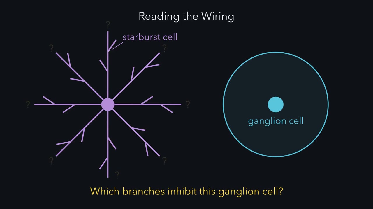
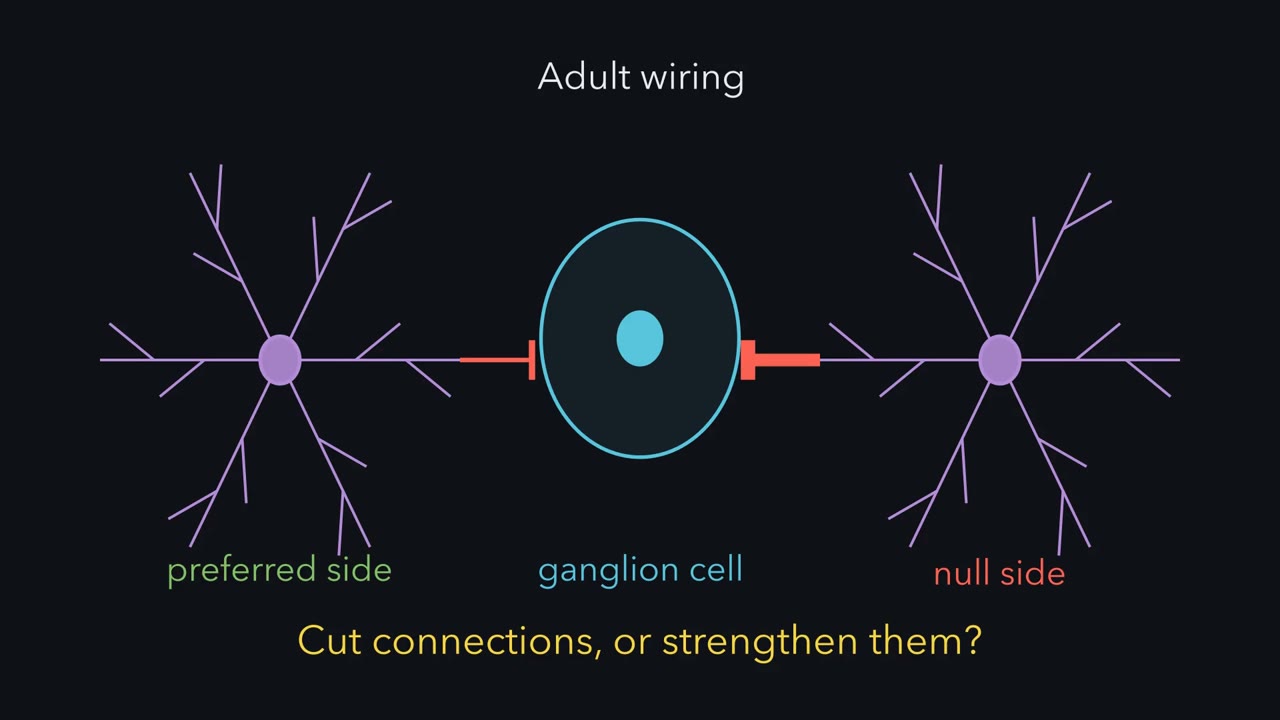
They also inferred that the veto is local, working in small subunits across the field. Here is a simple sketch. To cancel excitation, inhibition must overlap it in time. Null motion should start the inhibition early, so it should come from where null motion starts. Call that the null side. Preferred motion reaches the excitation first. The inhibition lags behind, so the excitation gets a head start. Barlow and Levick were tentative about which cells supply it. Most of it turned out to come from starburst amacrine cells. Its dendrites radiate like the spokes of a wheel. Each dendrite is its own small direction detector. It is most active when motion runs outward, from cell body to tip.
Take a ganglion cell that prefers rightward motion. It is wired mainly to starburst branches that point left, its null direction. Leftward motion runs outward along those branches. Their inhibition is strongest, and the ganglion cell stays quiet. Rightward motion runs inward along the same branches. They release little, so the excitation wins.
So remember the sign. The strongest inhibition comes from dendrites that prefer roughly the direction the ganglion cell ignores.
A cell in the eye fires to a spot moving left and stays quiet for the same spot moving right. Which neuron makes the difference?
Continue in the lesson: Computing Motion in the Retina →
2:50 · Free, with captions and a transcript. The lesson has the full text, the questions and the sources.
More films from this lesson
How this film was made. Narration: an AI narrator voice (Kokoro, af_heart) reading narration written from this lesson's text. Animation: AI-generated in code (Manim) by Claude, a schematic with no drawn molecular structures. Captions come from the narration script. See something wrong? Report it.