Biochemistry · Skim version
Techniques in Protein Biochemistry: Purify It, Then Prove It
Read the full lesson → Skim the whole course →
The guiding idea
Pull one protein out of a soup of ten thousand others — then prove you got it.
How do you pull one protein out of a soup of ten thousand others — and then prove you got it?
From the film
- An assay tracks the target through every purification step.
- Specific activity rises when a step retains a larger fraction of target activity than total protein.
- Yield tracks retained activity, while fold-purification tracks enrichment from the start.
- Choose a method using a property that distinguishes the target from contaminants.
-
Find it: the assay and the unit
-
Judge it: specific activity, yield and fold
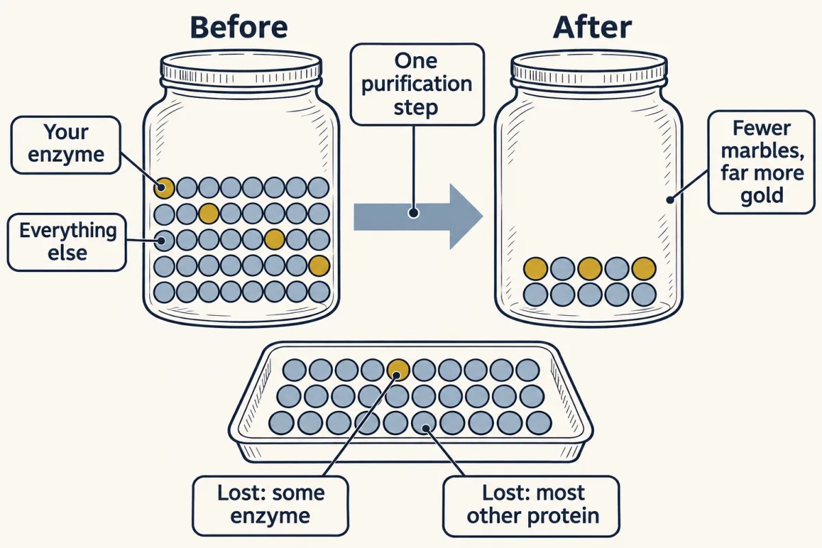
One step, counted. It kept 3 of 4 enzyme marbles (75%) but only 10 of 40 marbles overall (25%), so the gold fraction rose 0.75 ÷ 0.25 = 3-fold. Losing some enzyme is fine if you lose far more of everything else. AI-generated illustration -
Enrich it: one property per row
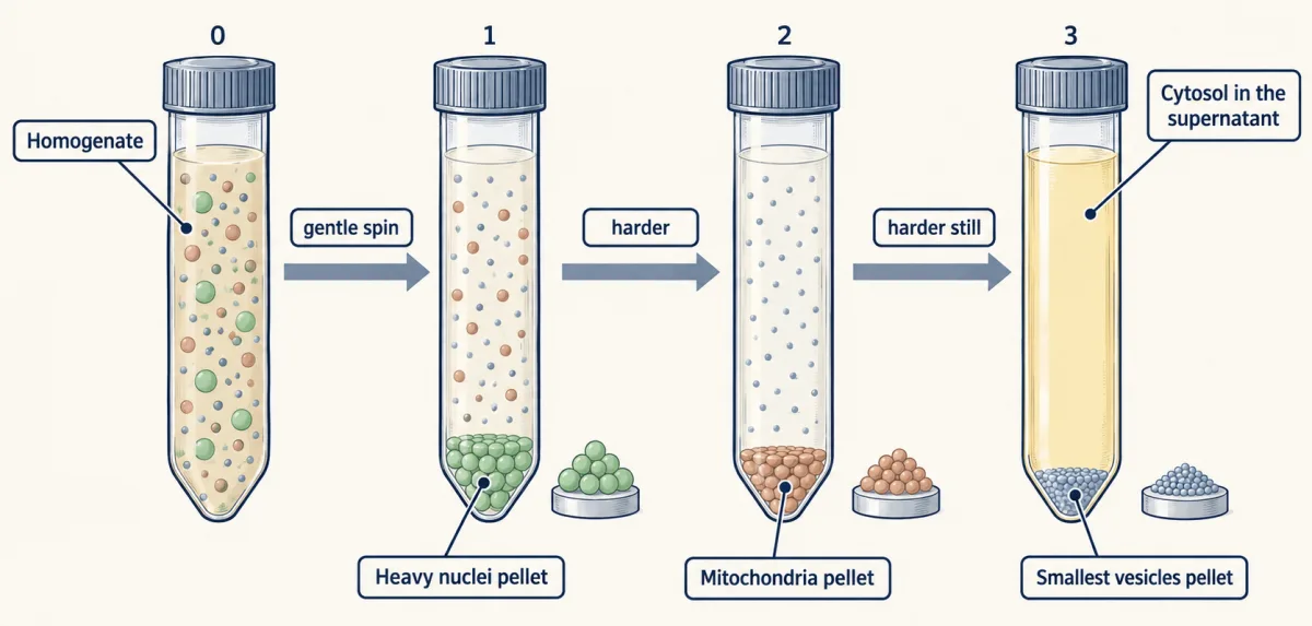
Sedimentation depends on size and density. After each spin the supernatant moves to a fresh tube and the pellet is set aside. The fractions are enriched, not pure. AI-generated illustration 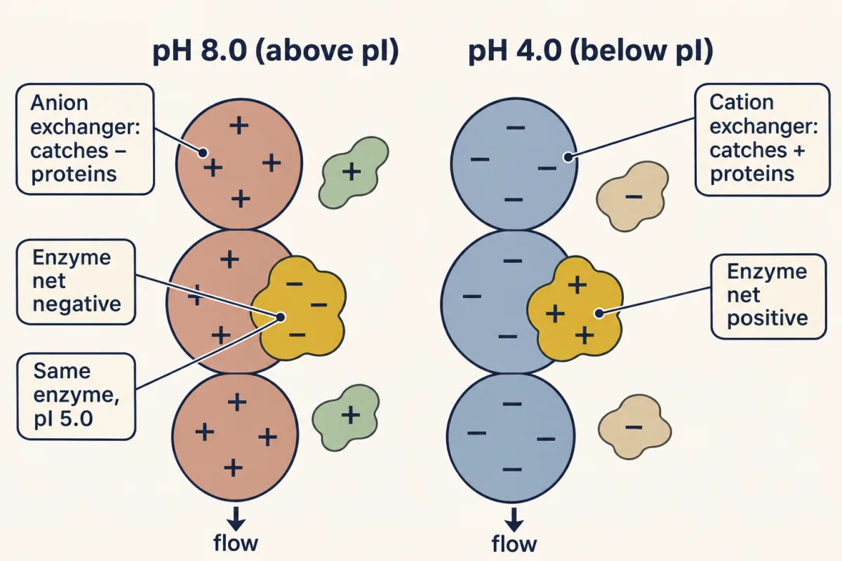
Name the resin by what it catches. The same enzyme (pI 5.0) is net negative at pH 8.0 and sticks to an anion exchanger, but is net positive at pH 4.0 and sticks to a cation exchanger. AI-generated illustration -
Know when to stop
-
Prove it: three questions
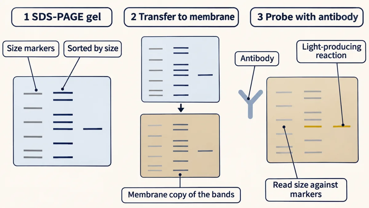
Gel, membrane, antibody: the three steps of a western blot. The membrane is an exact copy of the gel. A validated antibody supports identity, and the band's position against the size markers estimates its size. AI-generated illustration -
What each method answers
-
Going deeper
Where this lesson fits
Before thisReview first
- Amino Acids: How One Side Chain Decides EverythingThis course · Lesson 4
You need each side chain's charge and pKa from there to predict a protein's pI and how it behaves on an ion-exchange column.
- Protein Three-Dimensional Structure: The Sequence Sets the ShapeThis course · Lesson 5
Folded shape and subunit structure explain why affinity binding works and why SDS unfolds chains to sort them by size.
After thisStudy next
- Enzyme Kinetics: The Michaelis–Menten ModelThis course · Lesson 8
The activity assay and units used here become initial rates and Michaelis–Menten parameters measured on the purified enzyme.
- Basic Concepts of Enzyme ActionThis course · Lesson 7
It explains what an enzyme's catalytic activity is, the very quantity this lesson counts to follow the protein through each step.
Suggested by an AI model from the lessons themselves, then checked. How we use AI