Cell Signaling · Skim version
Exosomes and Extracellular Vesicles: Signals Between Cells
Read the full lesson → Skim the whole course →
The guiding idea
B cells release vesicles that carry their peptide-loaded MHC molecules, and those vesicles switch on only the T cells that recognise that peptide. A scrap of membrane can carry a signal.
Cells release small membrane vesicles, and these extracellular vesicles can carry a signal from one cell to another, either as a ligand on their surface or as protein and RNA inside.
-
Vesicles can carry a signal: the clearest case
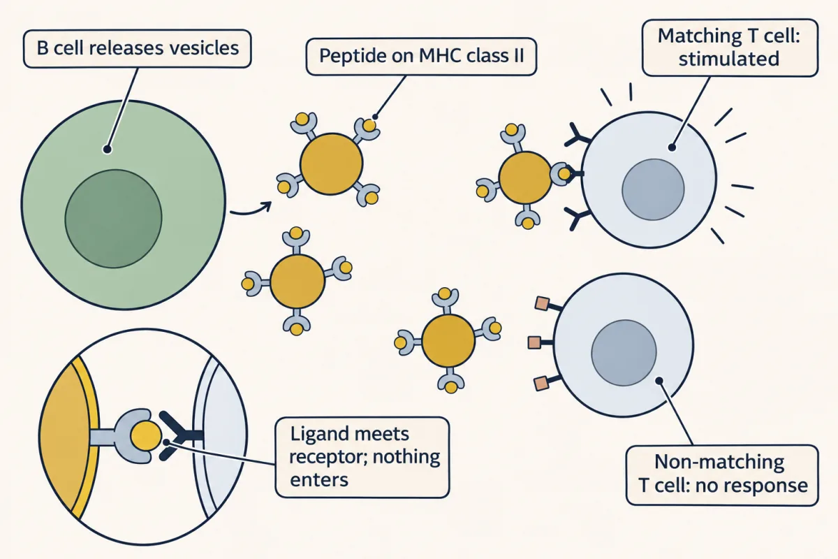
The clearest case: vesicles released by B cells carry peptide-loaded MHC class II on their surface and stimulate only T cells whose receptor recognises that peptide on that MHC molecule. The signal is a ligand meeting a receptor, so nothing has to enter the T cell. AI-generated illustration -
Where the vesicles come from: one compartment, two exits
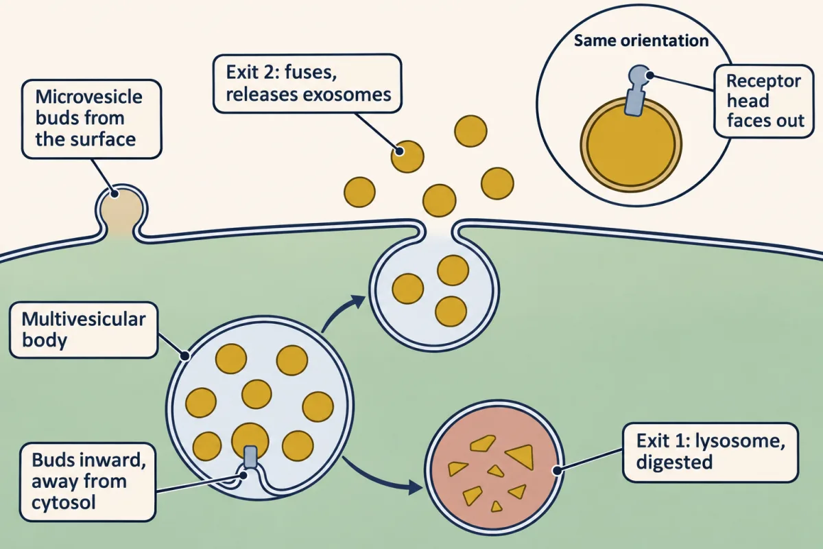
One compartment, two exits: a multivesicular body forms internal vesicles by budding inward, then either fuses with a lysosome, where they are digested, or fuses with the plasma membrane and releases them as exosomes. Because budding is away from the cytosol, a receptor on an exosome faces out as it did on the cell surface; microvesicles that bud straight from the surface have the same orientation. AI-generated illustration -
What is inside, and three ways it could act
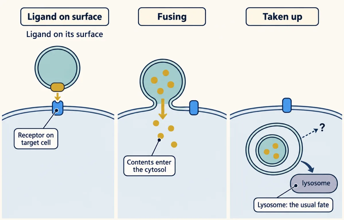
Three ways a vesicle could act on another cell. A ligand on its surface needs nothing to get in; fusion is the main proposed route that puts contents in the cytosol; uptake alone usually ends at the lysosome. AI-generated illustration -
How would you know? The pellet is defined by the centrifuge
-
Where the evidence is good, and where it is argued
-
Going deeper
Where this lesson fits
Before thisReview first
- Endocytosis and Signaling EndosomesThis course · Lesson 36
Multivesicular bodies and the endosome's route to the lysosome are used here, and exosomes are the second exit.
- Antigen Receptors and CheckpointsThis course · Lesson 29
The T-cell receptor recognising peptide on MHC is the receptor that B-cell vesicles switch on.
- A Field Guide to ReceptorsThis course · Lesson 4
A vesicle ligand acts as an ordinary receptor ligand, so you need how receptors read a ligand.
After thisStudy next
- Reverse Signaling: When the Ligand Is Also a ReceptorThis course · Lesson 39
It takes up a ligand held on another cell's membrane, as a vesicle surface ligand is, and asks which side gets the message.
Elsewhere on the siteFilms, simulations and method films in other courses
- Three Handoffs for a VesicleFilm · Cell Biology
Multivesicular body fusion with the plasma membrane is a vesicle targeting and fusion step.
- A Loop and a One-Way TripFilm · Cell Biology
Exosomes arise from endosomal membrane that is first taken in by endocytosis and sorted.
Suggested by an AI model from the lessons themselves, then checked. How we use AI