Physiology · Skim version
Cardiac Action Potentials and the ECG
Read the full lesson → Skim the whole course →
The guiding idea
A ventricular cell stays depolarised for about a fifth of a second. Why can't your heart cramp into a sustained contraction the way a skeletal muscle can?
Why does a ventricular cell stay depolarised for a fifth of a second, how does a pacemaker cell fire with no resting potential, and why is the electrocardiogram flat at the very moment every ventricular cell is depolarised?
-
One ventricular cell: a long plateau
-
Calcium in, more calcium out
-
Pacemaker cells have no resting potential
-
The wave spreads, pauses at the AV node, then races
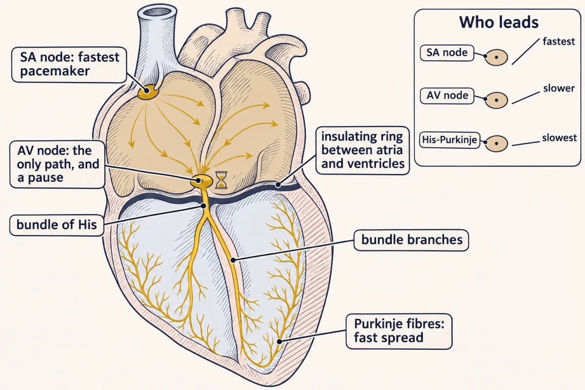
Excitation crosses the atria, pauses at the AV node, normally the only path through the insulating ring, then races through His and Purkinje fibres; slower pacemakers below stay silent because the SA node's wave fires them first. AI-generated illustration -
What the ECG records: cells that disagree
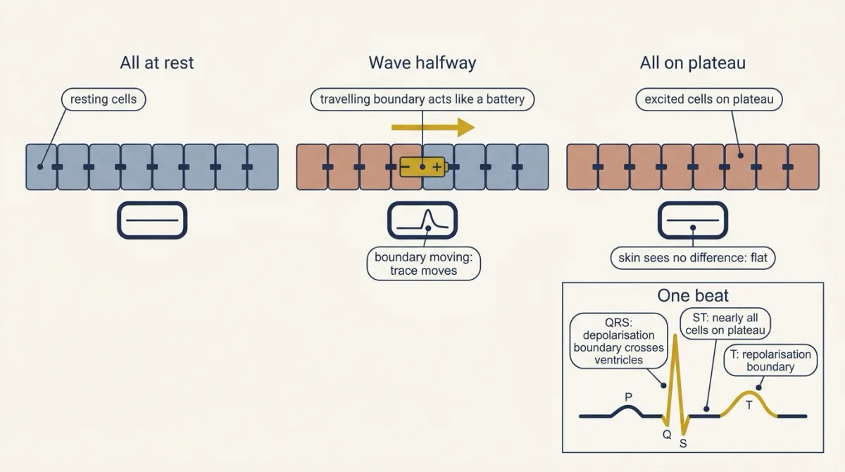
The ECG sees only boundaries between cells in different phases: when every ventricular cell is at rest, or every one is on its plateau, the trace is flat; it moves only while a boundary travels, which is why the ST segment is (nearly) flat although the ventricle is depolarised. AI-generated illustration -
AV block on the tracing
-
Potassium moves the resting potential and reshapes the ECG
-
Going deeper
Where this lesson fits
Before thisReview first
- Skeletal Muscle: Length, Load and SpeedThis course · Lesson 3
You need its twitch and tetanus summation to see why a long refractory period stops the heart from tetanising.
After thisStudy next
- The Cardiac Cycle and Pressure-Volume LoopsThis course · Lesson 6
It takes the calcium-driven contraction and relaxation from here and follows them through the pressure and volume of one beat.
- Cardiac Output and Venous ReturnThis course · Lesson 7
Heart rate and contractility, set here by pacemaker slope and PKA, become the heart side of cardiac output.
- Regulating Arterial PressureThis course · Lesson 10
Sympathetic and vagal control of the pacemaker and contractility here is the heart's part in reflex pressure control.
Elsewhere on the siteFilms, simulations and method films in other courses
- Patch clampMethod film
The plateau and funny currents are separated by recording currents through channels with a clamped voltage.
- Run the 1952 equationsSimulation · Neuroscience
Running the squid axon equations shows how sodium and potassium currents shape a spike, the base for the cardiac versions.
- Nernst calculatorSimulation · Cell Signaling
The Nernst calculator helps predict how raised potassium shifts the resting potential.
Suggested by an AI model from the lessons themselves, then checked. How we use AI