Cell Signaling · Skim version
Antigen Receptors and Checkpoints
Read the full lesson → Skim the whole course →
The guiding idea
The T-cell receptor has no enzyme and almost no tail. How does it tell the cell to kill?
How does a receptor with no enzyme and almost no tail switch on a T cell, and what keeps that cell from attacking healthy tissue?
-
The idea: one set of chains recognises, another set signals
-
The first minute: two phosphates, then three branches
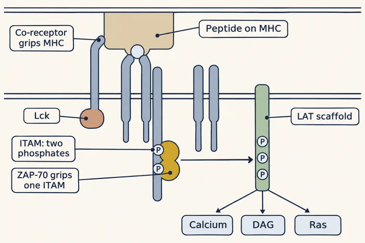
The receptor carries no enzyme. Lck, riding on a co-receptor that grips the same MHC protein, phosphorylates both tyrosines of an ITAM; ZAP-70 binds tightly only when both are phosphorylated, then phosphorylates the LAT scaffold, from which three branches leave: calcium, DAG and Ras. AI-generated illustration -
One signal is not enough: the second signal and two brakes
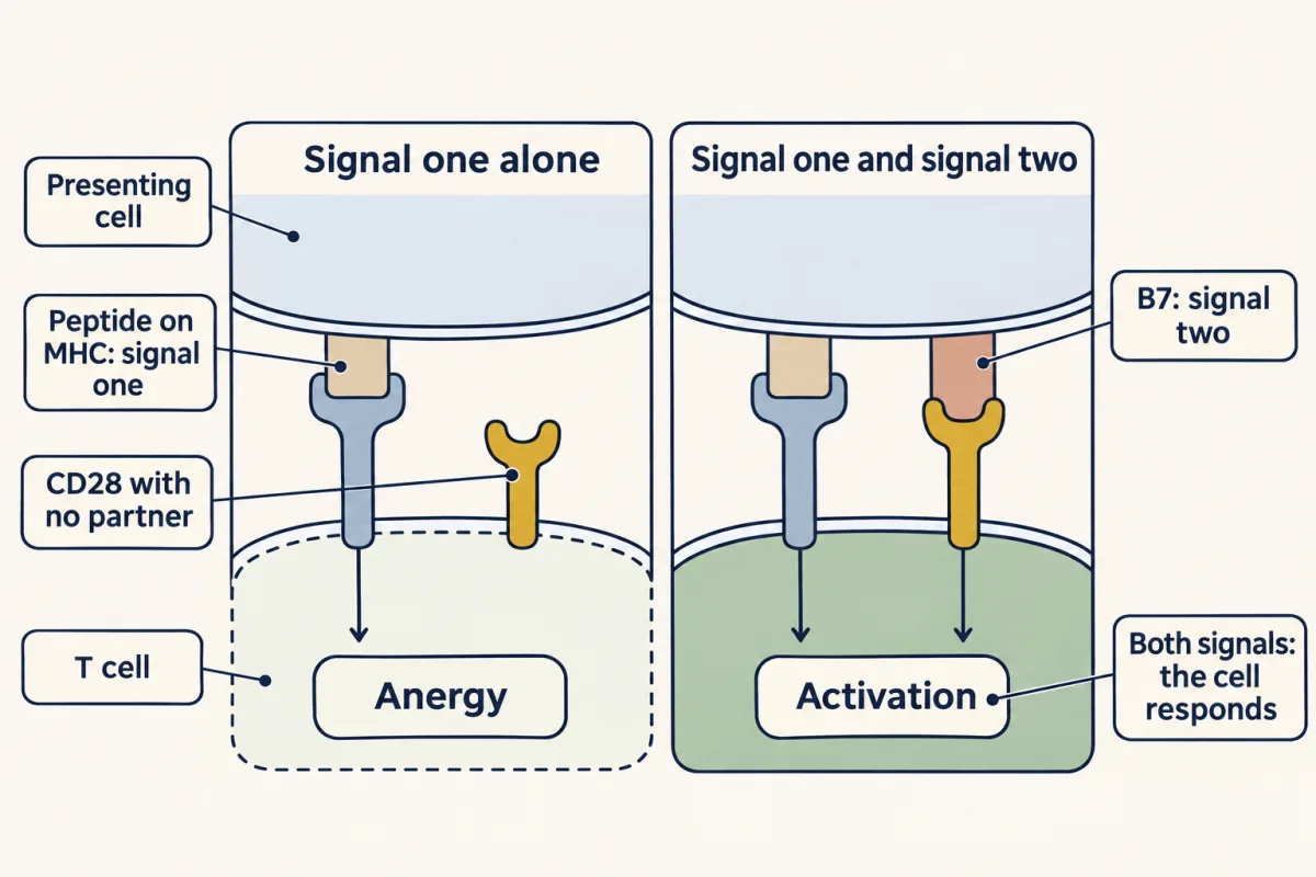
For a T cell meeting its antigen for the first time, signal one from the receptor is not trusted alone. CD28 must also bind a B7 protein, which presenting cells display once they have detected signs of infection. In the classic cultured-cell experiments, signal one alone left the cell in the lasting unresponsive state called anergy. AI-generated illustration 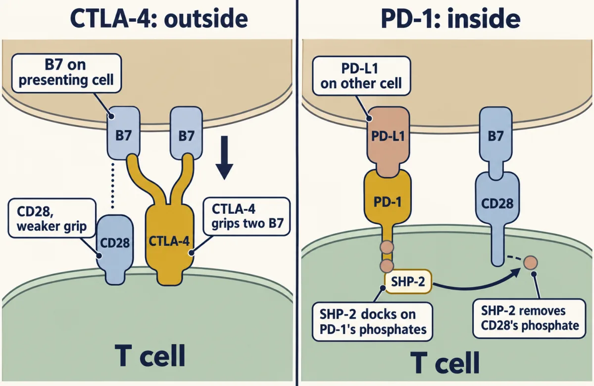
Two brakes, simplified. CTLA-4 binds B7 more tightly than CD28 does and can remove it from presenting cells, reducing co-stimulation; regulatory T cells use this route. PD-1 brakes from inside: once PD-L1 binds, its phosphorylated tail, mainly the ITSM motif, recruits the phosphatase SHP-2, which removes phosphates from activating signalling proteins such as CD28 and components of the antigen-receptor pathway. The tail phosphates themselves are the docking site, not the target. AI-generated illustration -
Releasing a brake to treat cancer
-
Building a receptor from parts
Where this lesson fits
Before thisReview first
- Phosphorylation: Kinases and PhosphatasesThis course · Lesson 6
Lck and ZAP-70 work by adding phosphates to tyrosines and CD45 and SHP-2 take them off, so you need that switch here.
- Protein Modules: SH2, SH3, PTB, PDZ and PHThis course · Lesson 9
ZAP-70 binds both phosphates of the ITAM through two SH2 domains, so you need to know how SH2 domains read phosphotyrosine.
- Adaptors, Docking Proteins and ScaffoldsThis course · Lesson 10
LAT is a membrane scaffold whose phosphorylated tyrosines recruit PLC-gamma, Grb2 and SOS, which you need to know first.
After thisStudy next
- The Shape of a Signal: Size, Duration, Frequency and PlaceThis course · Lesson 35
It takes up how the size and length of a signal decide the response, which bears on how T cells read signal strength.
- Drugs Against a PartThis course · Lesson 42
It carries on with how drugs are aimed at a single signalling part, as checkpoint antibodies are aimed at one receptor.
- Oncogenes: When the Switch SticksThis course · Lesson 41
It shows how stuck switches drive cancer, the disease that checkpoint blockade and CAR T cells treat.
Elsewhere on the siteFilms, simulations and method films in other courses
- Receptor binding and dose-response analysisMethod film
Receptor binding and dose-response analysis underlies how antibody and receptor affinity for antigen is measured.
- Receptors First, Antigen ChoosesFilm · Organismal Biology
It shows how each lymphocyte gets its one receptor shape, the antigen receptor this lesson then wires to signalling.
Suggested by an AI model from the lessons themselves, then checked. How we use AI