Developmental Neuroscience · Skim version
Two Places the Adult Brain Still Makes Neurons
Read the full lesson → Skim the whole course →
The guiding idea
Inject an adult rat with a label that marks newly made DNA. A month later a labelled cell sits in its hippocampus. Is it a new neuron, how would you know, and do you make them too?
A neuron counts as new only when the same cells carry two facts at once: a birth record, a label showing that their DNA was copied at a known time, so they were most likely born then or later, and a neuron identity.
From the film: Two Facts in One Cell
- A neuron counts as new only when the same cells carry a birth record and a neuron identity.
- A label such as BrdU dates the DNA, so a labelled neuron was most likely born then or later; controls are needed because DNA repair can also label a cell.
- A young-neuron marker such as doublecortin shows a cell's state, not when it was born.
From the film: Who Makes Them, and How Many Are Kept
- Adult rodents make neurons in two places, the subventricular zone and the dentate gyrus, where the rate falls steeply with age.
- Astrocyte-like stem cells make the neurons; after antimitotic treatment in mice, surviving B cells regenerated C cells and then neuroblasts.
- More new neurons can mean more division or more survival; in the classic mouse studies, running acted mainly on division and enriched housing mainly on survival.
From the film: Do You Make Them Too?
- In people, the evidence that carries a birth record, such as bomb-test carbon-14 dating, points to some hippocampal neurogenesis.
- Stains for young neurons show a state, not a birth date, and studies using them disagree.
- How many new neurons people make, and what they do, is still open.
-
How you know a neuron is new
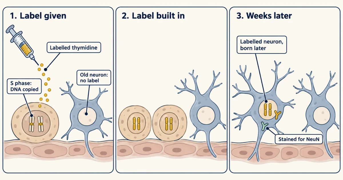
A labelled DNA building block (BrdU) is incorporated into DNA by cells that are copying it. A labelled neuron found weeks later most likely descends from a cell that was copying its DNA while the label was present, so it was born that day or later, although DNA repair can also label a cell, so controls are needed. Antibodies let the same cell also be stained as a neuron. AI-generated illustration -
Two places adult rodents keep making neurons
-
Which cell is the stem cell?
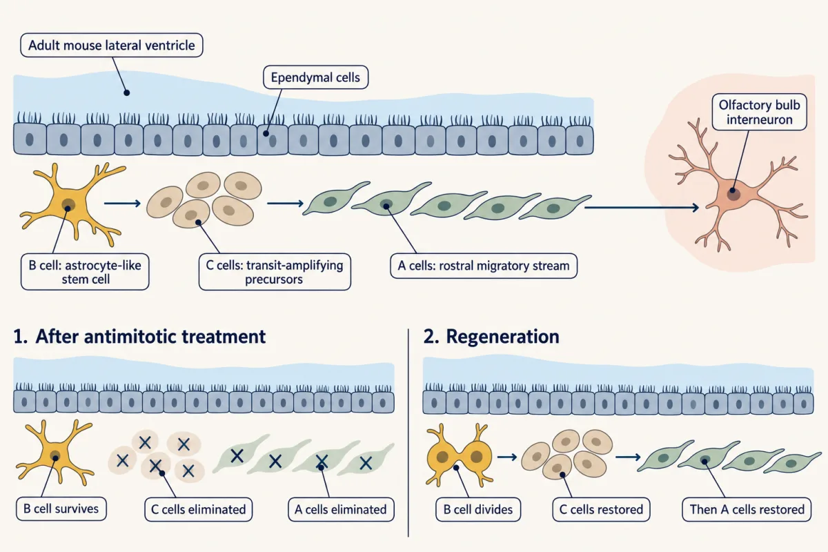
In the adult mouse subventricular zone, astrocyte-like B stem cells generate transit-amplifying C cells, then A-cell neuroblasts. Neuroblasts migrate along the rostral migratory stream to the olfactory bulb and become interneurons. After antimitotic treatment removed C cells and most A cells, surviving B cells divided and regenerated C cells followed by neuroblasts. AI-generated illustration -
Born is not the same as kept
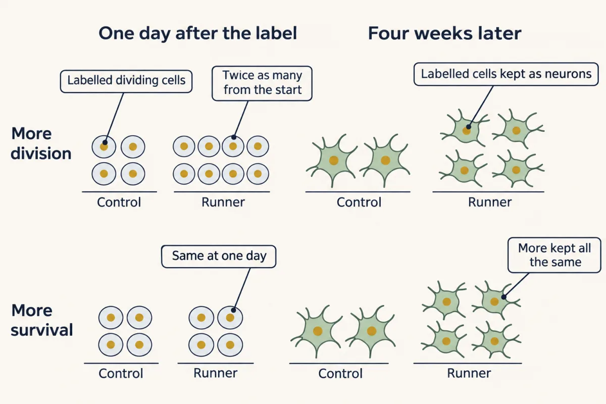
More new neurons can come from two different steps. Counting labelled cells one day after the dose indexes division; counting cells that carry both the label and a neuron protein weeks later counts those kept as neurons. Only the pair of counts tells the two apart, so a study reporting more new neurons has to say which step changed. Each row is a hypothetical outcome; in the classic studies, running acted mainly on division and enriched housing mainly on survival. AI-generated illustration -
New neurons join working circuits
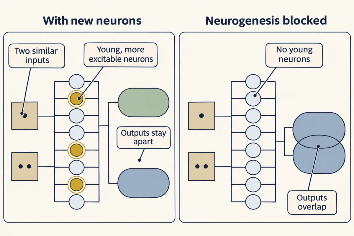
A leading hypothesis, not a settled result: a small, excitable young population helps the dentate gyrus give two similar inputs different outputs, the job called pattern separation. Mice in which adult neurogenesis was blocked did worse at distinguishing nearby locations. AI-generated illustration -
New neurons in the adult human brain
-
Putting it together
-
Going deeper
Where this lesson fits
Before thisReview first
- The Ventricular Zone: Radial Glia as Stem CellsThis course · Lesson 13
Adult stem cells here are astrocyte-like cells that resemble the radial glia introduced there as embryonic neural stem cells.
- Tangential Migration: Interneurons and the Long Way RoundThis course · Lesson 19
You need the chain migration of young neurons from there to follow the rostral migratory stream to the olfactory bulb.
- Too Many Neurons: Naturally Occurring Cell DeathThis course · Lesson 28
The idea of making too many neurons and keeping only some is used here for the early loss of newborn cells.
After thisStudy next
- Brains in a Dish: Stem Cells and OrganoidsThis course · Lesson 51
It takes up stem-cell-derived human neurons, a route to study human neurogenesis that cannot be done with birth labels.
Elsewhere on the siteFilms, simulations and method films in other courses
- How a Stem-Cell Pool Persists While Its Clones Come and GoFilm · Cell Biology
Shows how a stem-cell pool persists when identified by what its descendants do, not by a marker alone.
- Fate, Not SpeedFilm · Cell Biology
Shows how only dividing basal cells renew a tissue, a stem-cell fate question like this lesson's.
Suggested by an AI model from the lessons themselves, then checked. How we use AI