Neuroscience · Skim version
Synaptic Transmission and Quantal Release
Read the full lesson → Skim the whole course →
The guiding idea
A resting frog muscle fibre shows small voltage blips at its endplate with no nerve impulse at all. Those blips are the units of the evoked response at this junction.
Every evoked response at the frog neuromuscular junction is a whole number of packets, so the response is roughly count × size.
From the film: Blips from a Silent Nerve
- A nerve passes a signal to a muscle fibre in a relay: terminal impulse, calcium entry, vesicle release of acetylcholine, then receptor channels opening.
- A resting frog muscle fibre shows small blips with no nerve impulse, which Fatt and Katz attributed to acetylcholine released spontaneously.
- The miniature is the unit from which the evoked response at this junction is built.
From the film: Turn Release Down to See the Packets
- In normal calcium the units merge into one smooth response, so release has to be turned down to see them.
- In low calcium and high magnesium, responses cluster near whole-number multiples of the miniature, with failures at zero.
- Each response is roughly count times size, so when transmission changes, ask whether the count, the size or both changed.
From the film: Counting with Zeros
- The mean quantal content can be found two ways: average response divided by packet size, or from the fraction of failures.
- The failure method never uses packet size, so agreement between the two supports evoked and spontaneous packets being the same units.
- Low calcium makes failures common enough to count, which the mathematics needs.
From the film: Calcium Sets the Count
- Calcium entering through voltage-gated channels binds synaptotagmin and triggers SNARE-driven fusion.
- On the steep part of the curve release rises roughly as the fourth power of calcium, so a modest loss of calcium entry can cut release sharply.
- A slope near four is a reading of one curve, not a count of binding sites.
From the film: The Size of a Packet and the Margin of Safety
- Packet size depends on how much transmitter a vesicle holds and how many receptors face it.
- Each added packet moves the voltage less than the one before, so big responses fall short of count times size.
- Fewer receptors make each packet smaller while the count, set by the terminal and calcium, stays the same.
From the film: Count or Size? Two Weaknesses and a Synapse with No Packets
- In myasthenia gravis antibodies against acetylcholine receptors shrink the packet size, and in Lambert-Eaton syndrome antibodies against calcium channels cut the count.
- A smaller size with a normal count points to receptors or vesicle filling, and a lower count with a normal size points to release from the terminal.
- Electrical synapses pass current through gap junctions with no packets, so there is no quantal count or size.
-
The relay and the blips
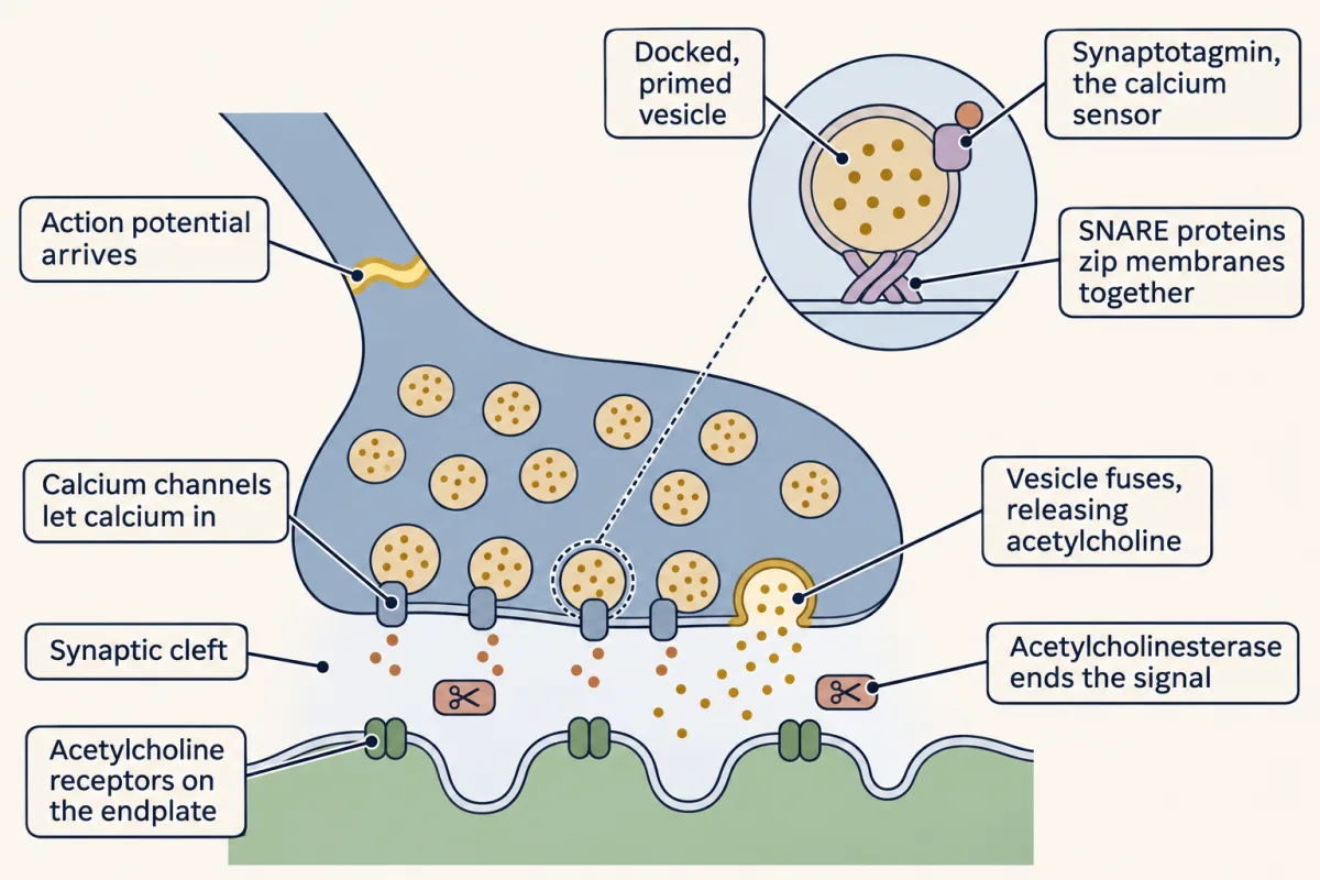
At the neuromuscular junction, calcium entry triggers vesicle fusion, released acetylcholine opens receptors on the endplate, and acetylcholinesterase ends its action. AI-generated illustration -
Packets: turning release down
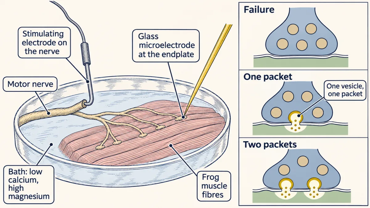
With release cut to a trickle by low calcium and high magnesium, each impulse released nothing, one packet or a few, so del Castillo and Katz could count the packets. AI-generated illustration -
Counting quanta you cannot see
-
Where the count comes from: calcium
-
Where the size comes from: receptors and the balance point
-
Count or size? When the safety factor fails
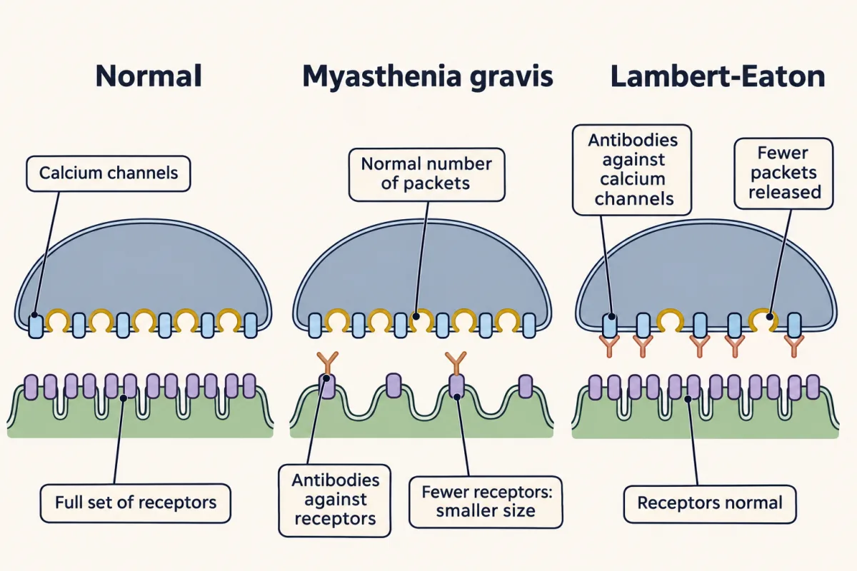
Two weaknesses from opposite sides: in myasthenia gravis antibodies remove receptors, so each packet is smaller, and in Lambert-Eaton syndrome antibodies against calcium channels cut the count of packets released. AI-generated illustration -
Going deeper
Where this lesson fits
Before thisReview first
- The Resting Membrane and Passive SignalsThis course · Lesson 3
You need the weighted-average balance point of conductances from there to follow how opening receptor channels depolarizes the endplate.
- The Action Potential and ConductionThis course · Lesson 4
The impulse that arrives at the terminal and opens calcium channels is described there, and this lesson starts from it.
After thisStudy next
- Transmitter Systems and DrugsThis course · Lesson 6
It takes the receptor and clearance steps here and asks how other transmitters and drugs change the size of a response.
- Synaptic PlasticityThis course · Lesson 8
It uses the count-times-size rule to ask whether changes in release come from the count or the size.
- Integration, Inhibition and NeuromodulationThis course · Lesson 7
It uses the postsynaptic conductance and balance-point idea to show how inhibition and summation shape the response.
Suggested by an AI model from the lessons themselves, then checked. How we use AI