Sensory Systems · Skim version
Sensory Neurons by Their Genes: the Atlas Era
Read the full lesson → Skim the whole course →
The guiding idea
Single-cell sequencing split the touch, pain and itch neurons of the dorsal root ganglion into many molecular kinds, far finer than C, Aδ and Aβ fibres. When does one of those clusters earn the name cell type?
Single-cell sequencing refined the broad fibre classes of mouse dorsal root ganglion neurons into many molecular subtypes: eleven classes in 2015, seventeen in 2018 and eighteen harmonized subtypes across six species in 2024.
From the film: From Cluster to Cell Type
- Atlas cluster counts depend on the data set, resolution, preparation and cell state.
- A transcriptomic cluster is a candidate type, not a functional conclusion.
- Genetic access lets researchers connect a candidate cluster to anatomy or physiology.
- An independent anchor strengthens a cluster's claim to be a functional type.
From the film: When Identity Changes
- Subtype identity emerges as broad transcription factor expression narrows during mouse development.
- After peripheral nerve injury, mouse sensory neurons can suppress subtype markers and enter a shared injury state.
- Subtype expression returns over a time course associated with axonal regrowth; timing alone does not prove cause.
- Mouse markers and cell classes do not automatically transfer to human sensory neurons.
-
What each atlas actually counted
-
From a candidate cluster to a functional test
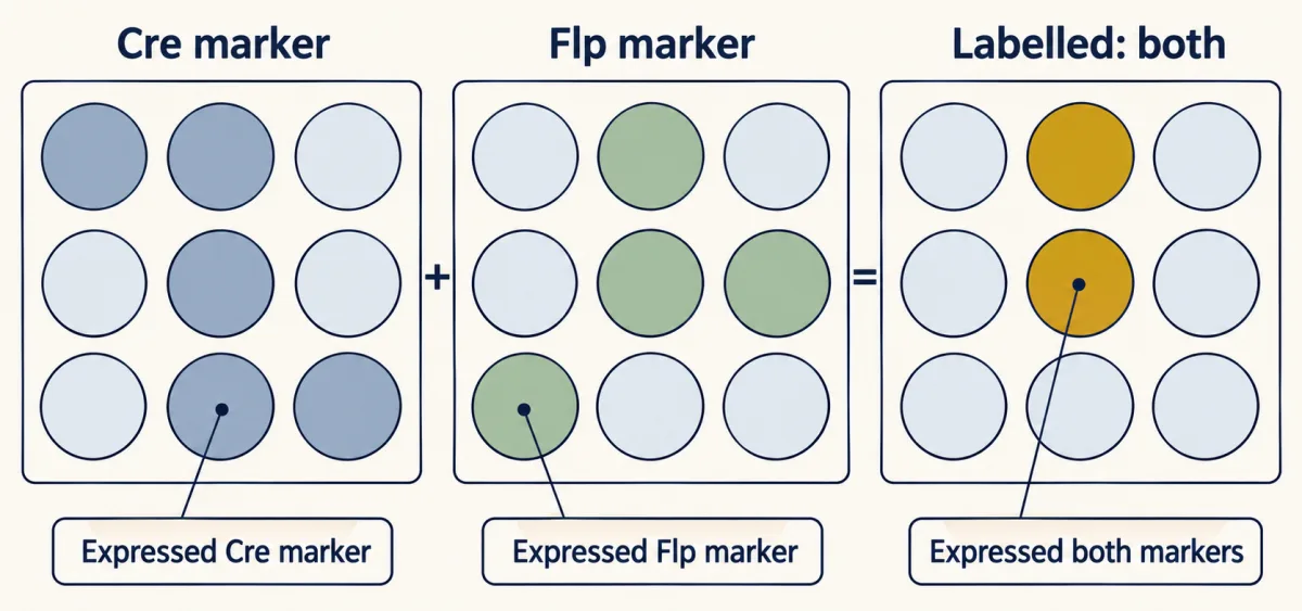
An intersectional strategy needs both Cre and Flp, so only cells that expressed both markers are labelled. The grid is a made-up illustration of the logic, not data. AI-generated illustration 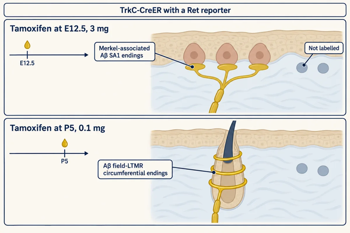
In mice carrying TrkC-CreER and a Ret reporter, 3 mg tamoxifen at E12.5 labelled Merkel-associated Aβ SA1 endings, whereas 0.1 mg at P5 favoured Aβ field-LTMR circumferential endings (Bai et al., 2015). Age and dose both differed. The drawings are schematic and show labelled ending types, not exact skin anatomy or the cells left unlabelled. A CreER label records expression history, not a fixed identity. AI-generated illustration -
The causal tests behind changing identity
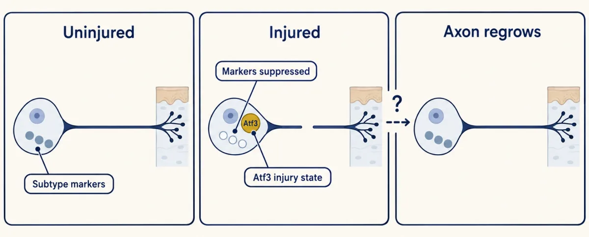
After peripheral nerve injury in mice, subtype-marker expression was suppressed and an Atf3-rich injury state appeared. Subtype expression re-emerged over a time course associated with axonal regrowth; this association alone does not show that reinnervation restores it. The symbols stand for expression states, not molecule counts, and an Atf3-rich cluster is a state existing neurons can leave, not a new type. AI-generated illustration -
Human atlases: methods, markers and denominators
-
Going deeper
Where this lesson fits
Before thisReview first
- Five Ways to Turn a Stimulus into a CurrentThis course · Lesson 1
It explains how different sensory neurons turn a stimulus into current, which the molecular subtypes here are sorted around.
After thisStudy next
- The Low-Threshold Mechanoreceptors: One Touch, Several SignalsThis course · Lesson 4
It builds the full touch-neuron class table that this lesson only points to, using genetic access to separate the classes.
- Nociceptors and the Sodium Channels of PainThis course · Lesson 11
It applies the mouse-to-human marker problem to pain neurons and the drug targets they carry.
- Itch: A Sense of Its OwnThis course · Lesson 13
It follows the itch-linked clusters, such as NP3 and Mrgpra3 neurons, into tests of function.
Suggested by an AI model from the lessons themselves, then checked. How we use AI