Physiology · Skim version
The Cardiac Cycle and Pressure-Volume Loops
Read the full lesson → Skim the whole course →
The guiding idea
Plot ventricular pressure against volume and every heartbeat draws a loop. Which corner moves when the arteries tighten?
Plot the pressure inside the left ventricle against its volume and each heartbeat draws a closed loop: its width is the stroke volume and its area is the stroke work.
-
One beat, four valve events
-
Fold time away and the beat becomes a loop
-
The ventricle’s line: a stiff line at end-systole, a soft curve at end-diastole
-
The arteries draw the second line
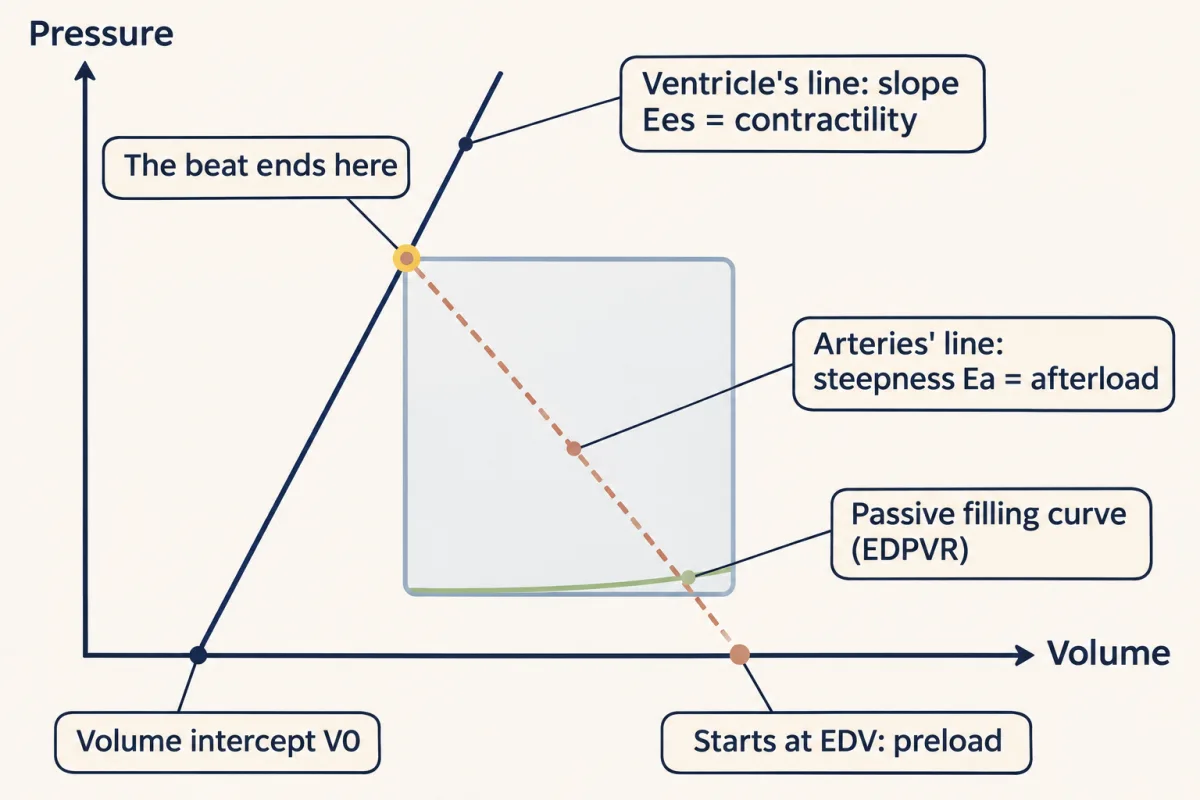
Each beat ends where the ventricle's line (slope Ees, contractility) meets the arteries' line (starting at EDV, steepness Ea, afterload); that crossing fixes end-systolic volume and pressure, and so the stroke volume. AI-generated illustration -
Preload, afterload and contractility each move one line
-
Ejection fraction is not contractility
-
Loops in disease: which line has moved?
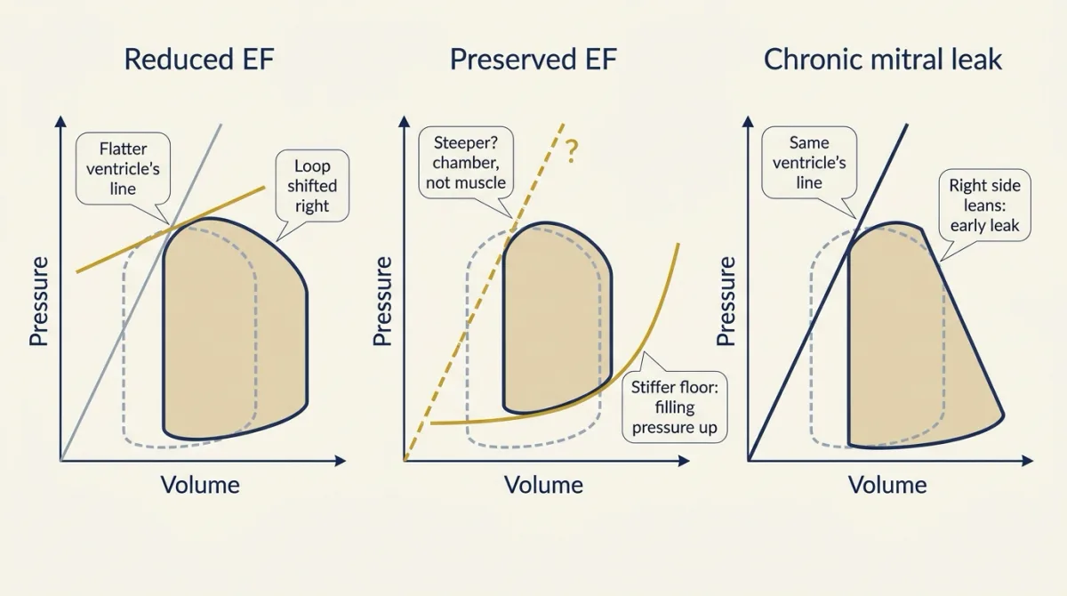
Disease moves different lines: reduced EF shifts the loop right and usually flattens the ventricle's line, preserved EF stiffens the filling floor (any steeper end-systolic line may reflect the chamber, not the muscle), and a mitral leak enlarges the loop with the ventricle's line unchanged, so EF can look normal. AI-generated illustration -
What a beat costs
-
Going deeper
Where this lesson fits
Before thisReview first
- Skeletal Muscle: Length, Load and SpeedThis course · Lesson 3
You need length, load and work from there to read the ventricle's end-systolic line and its stroke work here.
- Body Water and Its CompartmentsThis course · Lesson 1
Blood volume and its share of body water set the filling volume that this lesson takes as the starting point.
After thisStudy next
- Cardiac Output and Venous ReturnThis course · Lesson 7
It takes the loop's filling side and arterial load further, to see what sets cardiac output.
- Regulating Arterial PressureThis course · Lesson 10
It uses the arterial line (Ea) and the ventricle's line (Ees) to see how reflexes change pressure.
- Exercise: The Whole Body at WorkThis course · Lesson 30
It uses preload, afterload and contractility changes on the loop to explain how stroke volume rises in exercise.
Suggested by an AI model from the lessons themselves, then checked. How we use AI