Physiology · Skim version
The Ovarian Cycle and Fertility
Read the full lesson → Skim the whole course →
The guiding idea
In one 1984 study, single progesterone samples from normally cycling women in the mid-to-late luteal phase ranged from 2.3 to 40.1 ng per mL. What does one sample tell you?
How does an ovary that starts a whole cohort of follicles growing each cycle end up releasing one oocyte, at a predictable time after a hormone surge?
-
Most follicles die; one is chosen
-
Two cells, two gonadotropins, one estrogen
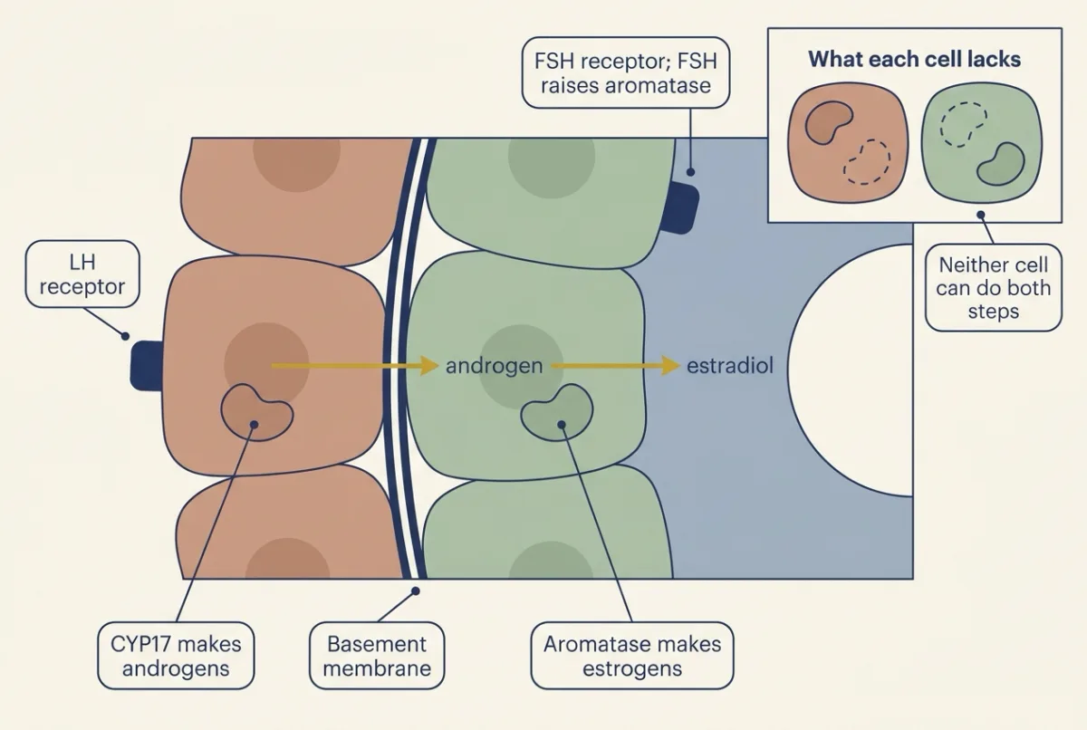
In the two-cell model the follicle makes estradiol as a two-station assembly: the theca cell makes androgen under LH, the granulosa cell aromatises it under FSH, and neither does much of both steps on its own, so the basement membrane is crossed in the middle of the pathway. AI-generated illustration -
From brake to accelerator: the switch to positive feedback
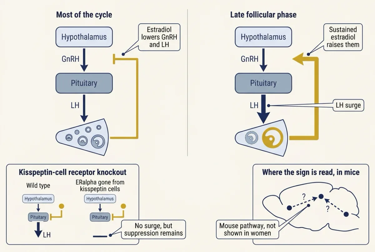
The same estradiol signal on the same axis reverses sign once it has stayed high for long enough; the mouse work tying the surge to kisspeptin cells is drawn dashed because in women the surge circuitry is less well localised. AI-generated illustration -
Ovulation and a corpus luteum that pulses
-
Reading the cycle’s hormone curves
Where this lesson fits
Before thisReview first
- Endocrine Axes and How to Test ThemThis course · Lesson 2
You need its feedback loops and the problem of reading one sample of a pulsatile hormone to follow this lesson.
After thisStudy next
- Pregnancy, Birth and LactationThis course · Lesson 29
Implantation, hCG rescue of the corpus luteum and the hormones of pregnancy continue directly from the luteal phase here.
Elsewhere on the siteFilms, simulations and method films in other courses
- Tap, Drain and a SensorFilm · Organismal Biology
It shows how a hormone reaches distant target cells with receptors, the basis of FSH and LH acting on follicle cells.
- A Loop That Keeps TimeFilm · Organismal Biology
It shows the hypothalamic pulses of GnRH driving pituitary LH and FSH, which set the ovarian cycle's rhythm.
Suggested by an AI model from the lessons themselves, then checked. How we use AI