Biochemistry · Skim version
Lipids: The Building Blocks That Refuse to Polymerize
Read the full lesson → Skim the whole course →
The guiding idea
Lipids make the barriers around every cell and the densest fuel in your body. One packing rule, how well the tails lie together, explains why a fat is solid or liquid and why a membrane is stiff or fluid.
Proteins, nucleic acids and polysaccharides are polymers; lipids are not.
-
Lipids break the rules
-
Fatty acids: why one bend melts a fat
-
Triacylglycerols: three tails on glycerol
-
Why lipids make bilayers (a separate rule)
-
Bilayers melt too: gel and fluid
-
Cholesterol, the buffer
-
The other job: fuel (a separate rule)
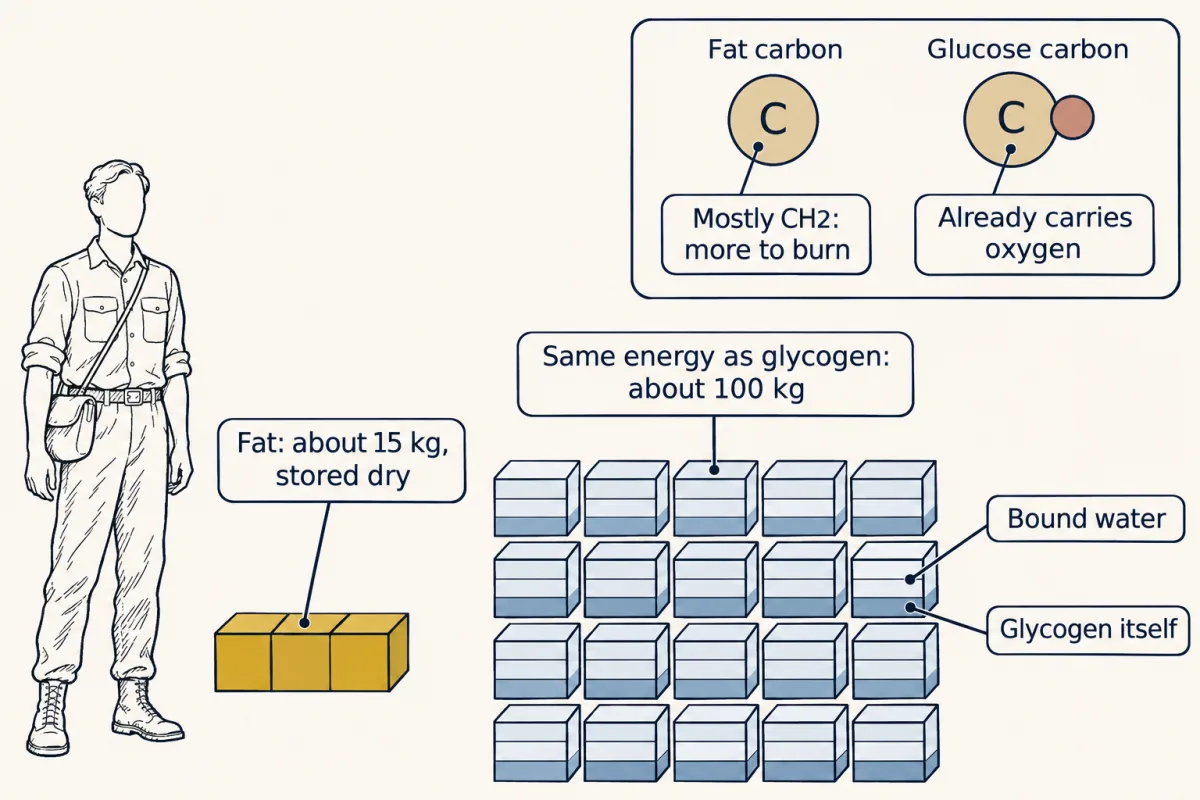
The same 135,000 kcal stored as fat weighs about 15 kg; stored as hydrated glycogen it would weigh about 100 kg. Two reasons, neither about packing: fat's carbon is more reduced, and fat is stored dry while glycogen holds about 2 g of water per gram. AI-generated illustration -
Membrane identity: head groups and leaflets
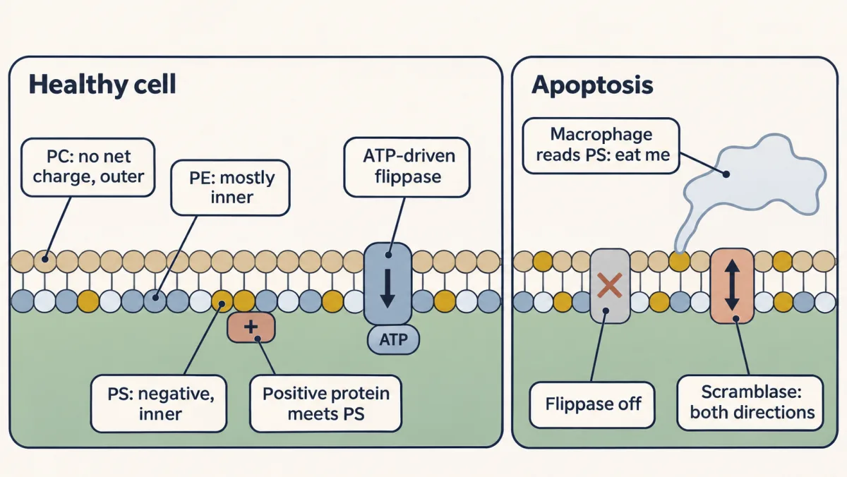
ATP-driven flippases keep phosphatidylserine (PS) on the inner leaflet of a healthy cell, where positively charged proteins meet it. In apoptosis the flippases shut off and scramblases let lipids cross both ways, so PS appears on the outer surface, where a macrophage reads it as an eat-me signal. AI-generated illustration -
Proteins at the membrane
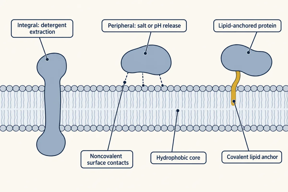
A protein can sit in the hydrophobic core, cling to the surface, or hang from a lipid anchor. A salt or pH change frees only the surface protein; core and most anchored proteins need detergent. AI-generated illustration -
Going deeper
Where this lesson fits
Before thisReview first
- Water, Weak Bonds, and the Generation of OrderThis course · Lesson 3
You need the hydrophobic effect and weak van der Waals contacts from there to follow why tails pack and bilayers form.
- Introduction to Biochemistry and Cell Biology: Compartments Create ControlThis course · Lesson 2
It introduces compartments and membranes as barriers, which this lesson explains at the level of the lipids themselves.
After thisStudy next
- Membranes: The Impermeable Sheet That Builds ItselfThis course · Lesson 12
It uses the bilayer assembly, fluidity and cholesterol buffering taught here to explain how membranes block ions and carry proteins.
- Fatty Acid Degradation: The β-Oxidation SpiralThis course · Lesson 25
It breaks down the reduced, anhydrous fat store described here, two carbons at a time, to harvest its energy.
- Fatty Acid Synthesis: Building the Chain Two Carbons at a TimeThis course · Lesson 26
It builds the saturated chains whose length and double bonds set the packing rule used here.
Elsewhere on the siteFilms, simulations and method films in other courses
- Set Up: The Blubber GloveFilm · Biology Together
Shows fat as insulation and a body store of energy, which links to fat as the dense fuel here.
Suggested by an AI model from the lessons themselves, then checked. How we use AI