Biochemistry · Skim version
Carbohydrates: One Bond Decides Fuel or Fiber
Read the full lesson → Skim the whole course →
The guiding idea
Closing the glucose ring makes one special carbon, and the direction of the bond it forms decides whether a polymer feeds you or holds a plant up.
Starch and cellulose are both pure glucose, yet you digest one and not the other.
From the film
- Starch and cellulose are both glucose polymers; their 1,4 bonds use opposite faces.
- Closing the glucose ring turns carbon one into the anomeric carbon, whose new group can point to either face.
- β puts that group on the same face as carbon six; α puts it on the opposite face.
- For glucose joined 1 to 4, β chains run straight into cellulose fibers; α chains coil into starch and glycogen.
- You digest starch because your amylases fit α-1,4; you make no cellulase to cut cellulose's β bonds.
-
Meet glucose: just enough to number it
-
Closing the ring makes one special carbon
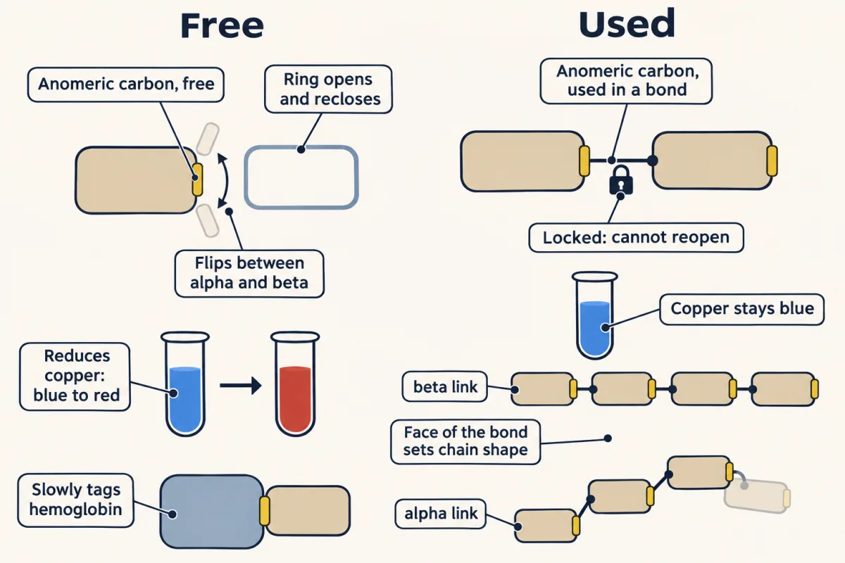
The lesson's one idea. Free, the anomeric carbon lets the ring reopen: the sugar flips between α and β, reduces copper and slowly tags proteins. Used in a bond, it is locked, and the face of that bond sets the shape of the chain. AI-generated illustration -
Naming the relationships
-
A free anomeric carbon can reopen
-
A used anomeric carbon: the glycosidic bond
-
Bond geometry sets polymer shape
-
Branching: more ends, faster fuel
-
The sugar coat: a second rule
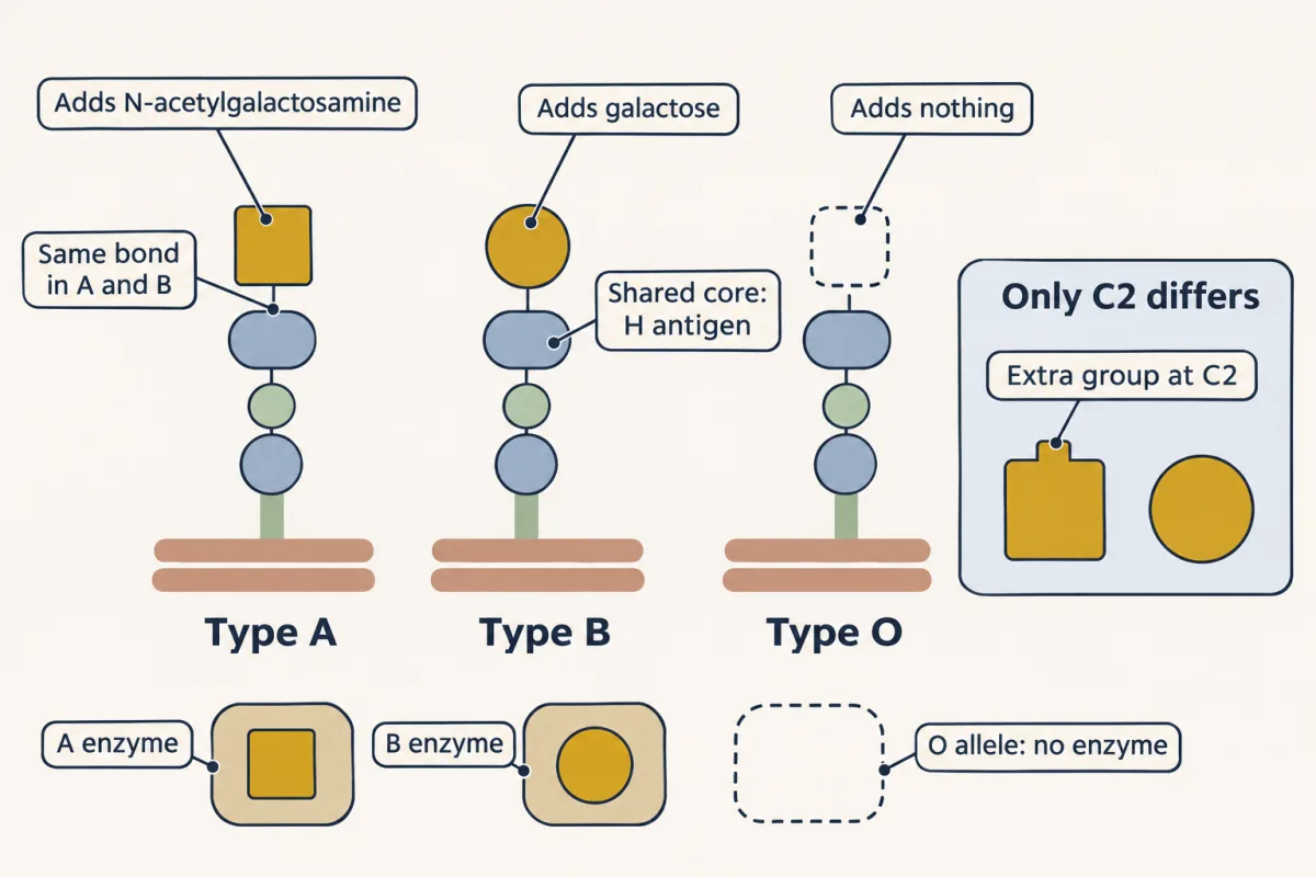
Your ABO type is the second rule at work: which sugar an inherited enzyme adds to the shared H antigen. The A enzyme adds N-acetylgalactosamine, the B enzyme galactose, by the same bond; the O allele adds nothing. The two sugars differ only at C2. AI-generated illustration 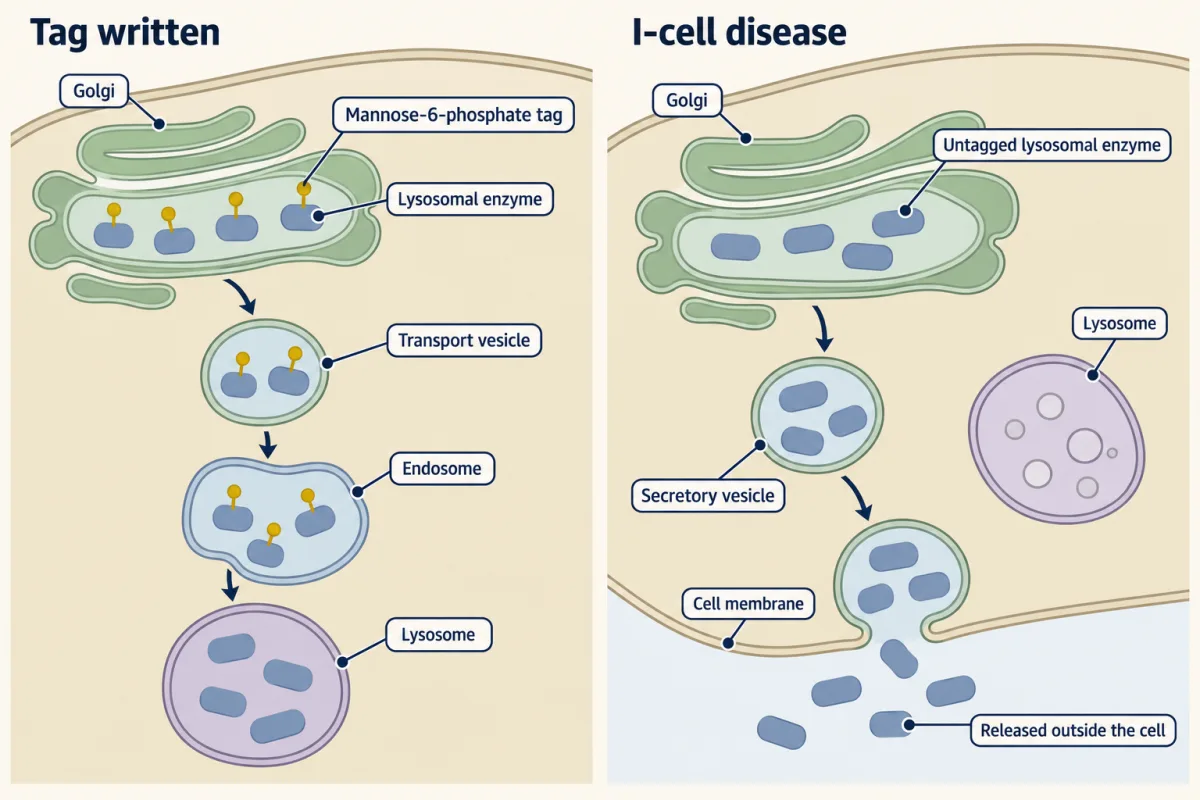
A mannose-6-phosphate tag is the address that sends enzymes to the lysosome. Lose the ability to write the tag and the enzymes are dumped into the blood instead, the lost-mail problem behind I-cell disease. AI-generated illustration -
Lectins read what the sugar code wrote
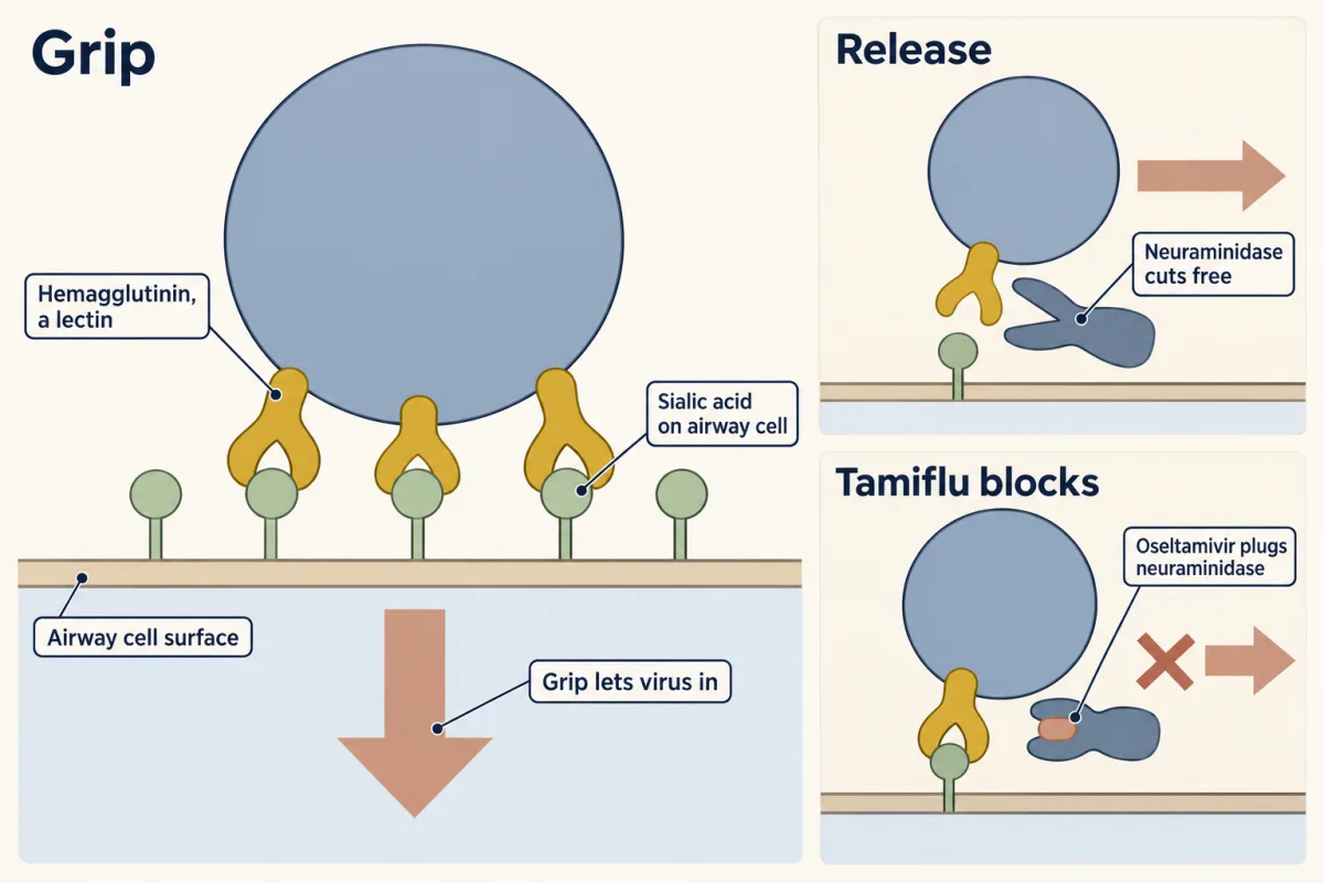
Influenza hemagglutinin, a lectin, binds terminal sialic acid on airway-cell glycans to attach before entry. Viral neuraminidase removes terminal sialic acids, helping new virions release. Tamiflu inhibits neuraminidase, reducing release and spread. AI-generated illustration -
The idea, with its scope
-
Going deeper
Where this lesson fits
Before thisReview first
- Water, Weak Bonds, and the Generation of OrderThis course · Lesson 3
You need hydrogen bonding and weak interactions from there to see how cellulose chains pack and why glucose dissolves.
- What Am I Made Of?Biology · High school
It introduces carbohydrates as polymers built from sugar monomers, which this lesson then explains in detail.
After thisStudy next
- Glycolysis: The Investment-and-Payoff PathwayThis course · Lesson 16
The free glucose ring you met here is the molecule this pathway breaks down.
- Digestion and Absorption: Turning a Meal into Molecules Your Cells Can UseThis course · Lesson 14
Amylases cutting α-1,4 bonds and lactase cutting β-1,4 bonds are followed here through the gut.
- Integration of Metabolism: How the Body Switches FuelsThis course · Lesson 30
Glycogen branching and fast mobilization from non-reducing ends are used here when the body switches fuels.
Suggested by an AI model from the lessons themselves, then checked. How we use AI