A Synapse That Does Not Wait
Read the transcript of “A Synapse That Does Not Wait”
Film transcript: A Synapse That Does Not Wait
Many irregular fibres start at type I hair cells, each wrapped in a cup-shaped ending called a calyx. Could this synapse help make them fast? A type I cell is flask-shaped, almost enclosed by its calyx. A type II cell is cylindrical, with small boutons. Calyx-only fibres, and many irregular fibres, come from each organ's central zone. Type I cells occur in both zones. That is a population pattern. A tempting shortcut says type I cells release no vesicles. Wrong. Both types release glutamate from vesicles. In sustained release, type I cells in mature mouse tissue released about ten times less than type II cells. Now a second route. Non-quantal transmission is a change in calyx voltage with no vesicle fusion and no gap junctions.
At a chemical synapse, a vesicle must fuse, glutamate must cross, and receptors must open. Each step adds delay. Pause and predict. Remove otoferlin, or a calcium channel that release needs. Can a type I cell still make its calyx fire?
Yes. Without either protein, type I cells still made the calyx fire, in mouse tissue in a dish. The test was whether spikes survive, not whether the signal is normal. How could that work? The cleft between the cell and its calyx is very narrow. When the hair cell depolarizes, more potassium current flows out into that thin space. Potassium slowly builds up there. Raise potassium in the cleft, and potassium's reversal potential at the calyx moves up. With the other conductances held fixed, this shift tends to depolarize the calyx. The current also makes the cleft itself positive, quickly. Less potassium leaves the calyx, or some enters, so it depolarizes.
We met potassium first, but in a model, the cleft potential acts first and potassium later. That order is a prediction. Keep the evidence apart. Mouse knockouts and young rat calyces are measurements. So are turtle recordings of both cells together, which found a slow coupling and a much faster one.
Two routes leave a type I cell. One waits for vesicles. One does not.
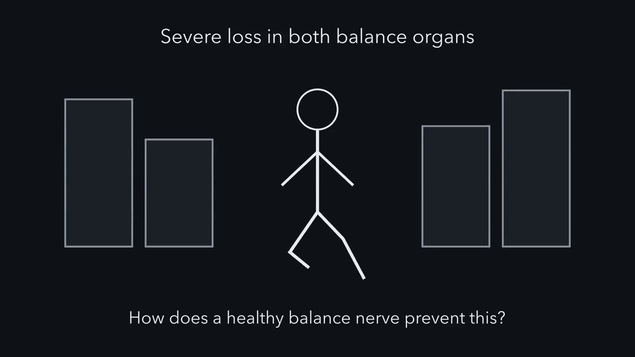
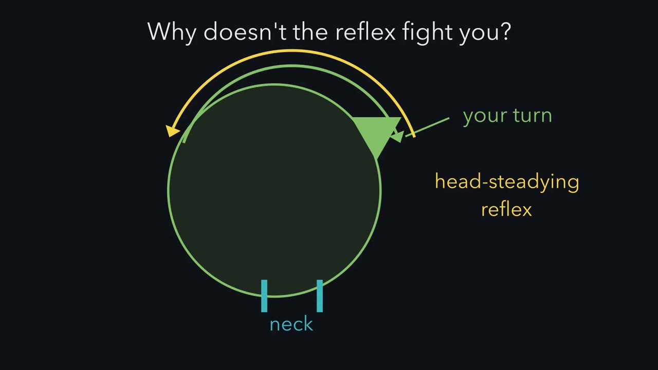
People with severe loss in both balance organs can see the world bounce as they walk. Yet in monkeys, head-motion neurons go largely quiet for a turn the animal makes itself, which may keep the head-steadying reflex from fighting it. How does one nerve report motion fast and precisely, yet let the brain set your own turns aside?
Continue in the lesson: Balance: Two Codes for Every Head Movement →
2:44 · Free, with captions and a transcript. The lesson has the full text, the questions and the sources.
More films from this lesson
How this film was made. Narration: an AI narrator voice (Kokoro, af_heart) reading narration written from this lesson's text. Animation: AI-generated in code (Manim) by Claude, a schematic with no drawn molecular structures. Captions come from the narration script. See something wrong? Report it.