Films · Developmental Neuroscience
Delete One, Count the Survivors
Read the transcript of “Delete One, Count the Survivors”
Film transcript: Delete One, Count the Survivors
If you remove one receptor from a mouse, which neurons disappear, and when? A knockout mouse is one in which a chosen gene has been disrupted. It makes no working protein, or no working form of one protein. Delete each neurotrophin or Trk gene, one at a time. The family sorts by which neurons are lost. Start with TrkA, and the sympathetic ganglion in the neck. Anne Fagan and colleagues counted its cells at several ages. At embryonic day 13.5, the count was normal. If TrkA is needed for survival, not for making the neurons, what should happen next?
The mutant fell steadily behind. At birth it had about half as many neurons as control ganglia. By postnatal day 9, nearly all of its principal neurons were gone. Over those embryonic ages, dividing cells were no fewer than in controls. Dying cells, with shrunken nuclei, increased. So the neurons were made, and then they died. Now the other pairs. Mice lacking NGF or TrkA lose sympathetic neurons. They also lose many small sensory neurons that sense pain and heat. Mice lacking BDNF lose vestibular neurons, which sense balance and head movement. They also lose many nodose-petrosal neurons, which carry signals from the heart, lungs and gut. Mice lacking NT-4 lose nodose-petrosal neurons too.
And mice lacking NT-3 or TrkC lose the sensory neurons that report where a limb is. These mice move and hold their limbs abnormally. In this grid, lose means fewer neurons. Usually part of a ganglion survives, but some groups are lost almost entirely. Each pair has its own pattern of loss. But the map has overlaps. NT-3 prefers TrkC, yet it also activates TrkA and TrkB. Should a mouse lacking NT-3 lose more neurons, or fewer, than one lacking TrkC?
Expect more. In many ganglia at the same age, mice lacking NT-3 lose more neurons than mice lacking TrkC. That fits NT-3 also acting through TrkA and TrkB. The size of the gap depends on the ganglion and age. Some populations also need more than one factor, at different stages. The takeaway: each factor and receptor pair has its own pattern of loss. In the sympathetic ganglion, neurons were made, then lost.
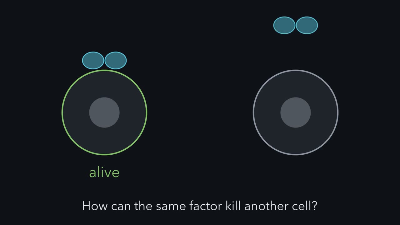
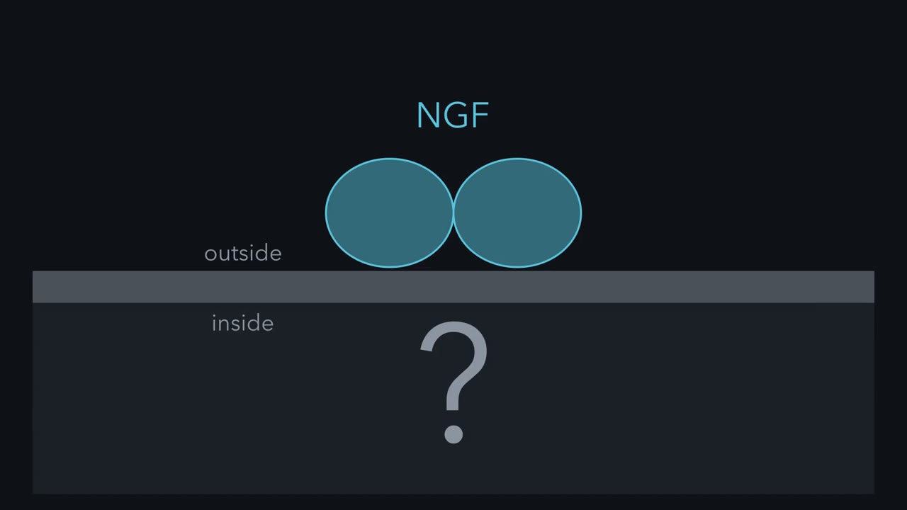
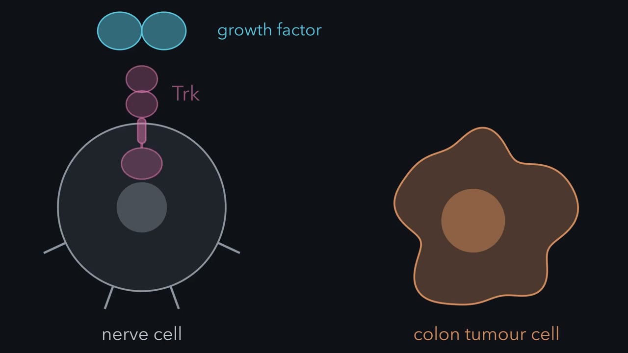
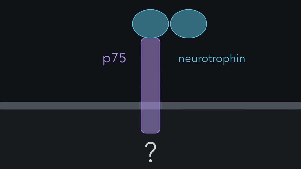
Many developing neurons live only if their target supplies enough of the right neurotrophin. How does the neuron read that factor, and how can the same factor sometimes kill?
Continue in the lesson: Neurotrophins, Trk Receptors and p75 →
3:03 · Free, with captions and a transcript. The lesson has the full text, the questions and the sources.
More films from this lesson
How this film was made. Narration: an AI narrator voice (Kokoro, af_heart) reading narration written from this lesson's text. Animation: AI-generated in code (Manim) by Claude, a schematic with no drawn molecular structures. Captions come from the narration script. See something wrong? Report it.