Films · Developmental Neuroscience
Whose Cells Built It?
Read the transcript of “Whose Cells Built It?”
Film transcript: Whose Cells Built It?
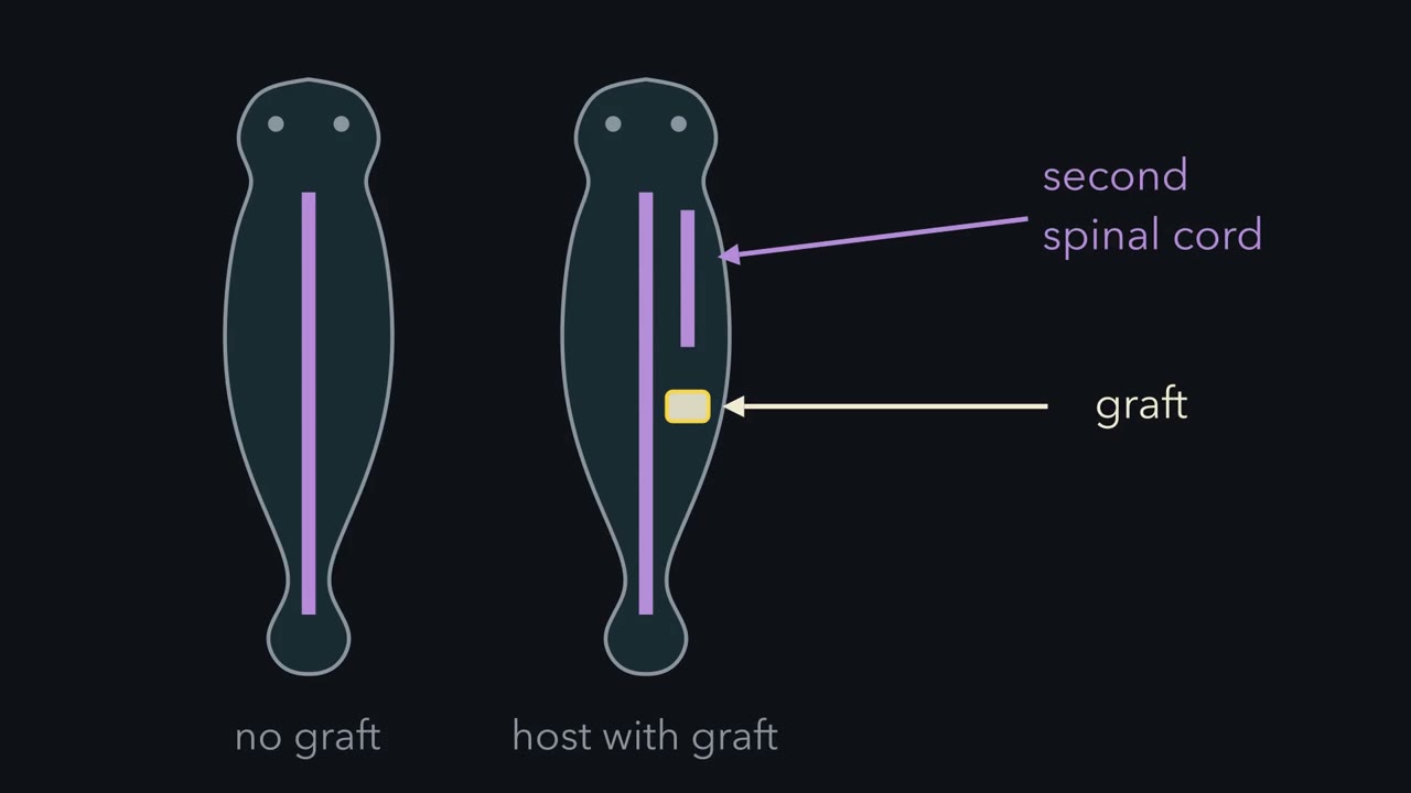
A dorsal lip moved to a new place can give a second body axis. But whose cells built it? Grafts within one species had given extra axial structures. But donor and host cells look alike. So, two readings for the new neural tube. Self-differentiation: the graft's cells develop by their own origin, and make the tube themselves. Or induction: the graft changes the fate of its neighbours, and host cells build the new tube. The solution was colour. Hilde Mangold did the test in Hans Spemann's laboratory, for her doctoral thesis. She used two newt species. Cells of one carry little pigment. Cells of the other carry pigment granules. The granules stay visible in sections, so colour shows which embryo a cell came from.
In 1921 and 1922, she cut a piece of dorsal lip from a pale gastrula. She placed it in the belly of a pigmented gastrula, in tissue that would normally become skin. Most embryos died. In the survivors, a second neural plate rose, folded, and closed into a second neural tube. Predict. If the graft simply grew, what colour is the new tube? If it changed its neighbours?
If the graft grew, the tube should be pale. If it changed its neighbours, the tube should be pigmented. In one embryo, most of the second tube was pigmented, with a narrow pale strip along its floor. In another, the whole tube was host tissue. The pale graft lay beneath it, as a notochord and nearby mesoderm.
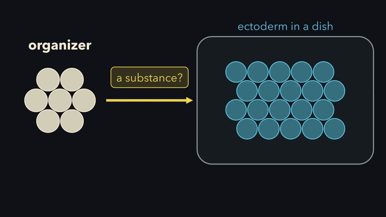
Host ectoderm that would have made skin had made a neural tube. For the tube, induction explained it. Induction is a change in the fate of one tissue, caused by a signal from another. The graft did three things. Its own cells became notochord. It recruited host mesoderm into somites, blocks beside the tube that form vertebrae and muscle. And it induced host ectoderm to become a neural tube. Hence the name, organizer. The second axis formed at the graft, not along the host's own back. The pattern followed the source. So the organizer decides where neural tissue forms. It does not yet say how: by a go signal, or by releasing a brake.
The responder must also be able to answer. That ability is competence. Wrap older and older ectoderm around early organizer. The response falls, and largely stops by the end of gastrulation. The control: late organizer with young ectoderm. If that still works, the loss is in the responder.
So host cells built the second neural tube, at the graft, while they could still answer.
How can a small graft make its host build a second spinal cord from cells meant for skin?
Continue in the lesson: The Organizer: Spemann and Mangold →
3:21 · Free, with captions and a transcript. The lesson has the full text, the questions and the sources.
More films from this lesson
How this film was made. Narration: an AI narrator voice (Kokoro, af_heart) reading narration written from this lesson's text. Animation: AI-generated in code (Manim) by Claude, a schematic with no drawn molecular structures. Captions come from the narration script. See something wrong? Report it.