Films · Developmental Neuroscience
Caught in the Act
Read the transcript of “Caught in the Act”
Film transcript: Caught in the Act
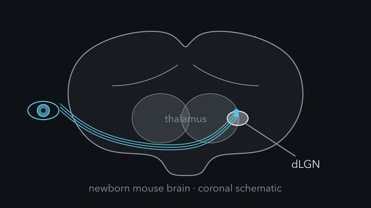
If microglia eat retinal inputs, can we catch them doing it? And do they choose what they eat? In 2012, Dorothy Schafer, in the Stevens laboratory, set out to look. Each eye got its own colored tracer at postnatal day four. A day later, they looked in the visual thalamus. A speck beside a microglial cell does not show it was eaten. So only label inside the cell counts. Retinal material sat inside microglial lysosomes, the cell's digestive compartments. A dye that glows only in acid agreed. The swallowed material was mainly presynaptic, from retinal terminals.
Eating had a clock. It was high at postnatal day five, and much lower by day nine. Eating also used a tag and a reader. Without complement receptor 3, the reader, or C3, the tag's source, microglia took up about half as much. Those mice later had more overlap between the eye territories, and extra retinal synapses. But half the eating remained. Complement is one route of several. Now the key question. Silence one eye with tetrodotoxin, which blocks sodium channels. Predict. If microglia remove less active inputs, whose material should they hold more of?
More of the silenced eye's material, relative to how much of that eye's input is there. Boost one eye with forskolin instead, and they take relatively more of the untreated eye's. The two drugs push in opposite directions. A drug that simply damaged axons would raise eating of its own eye both times.
Why relative to what is there? Near one microglial cell, one eye's input can far outnumber the other's. Say 90 units from one eye, and 10 from the other. An eater with no preference takes a tenth of each, so it holds 9 to 1. Now silence the minor eye, and let the eater take twice the fraction of it: two tenths instead of one tenth. It holds 2 units, still fewer than 9. So a fair test compares each eye with its own input, measured where the microglia are sampled. Now the limits. This compares whole eyes, not single synapses. It does not show how activity becomes a mark. And later imaging elsewhere suggests microglia often take bites, not whole synapses.
Here is the idea again. Relative activity biases how much material microglia take from each eye. Complement helps them take part of it. How the two connect is still open.
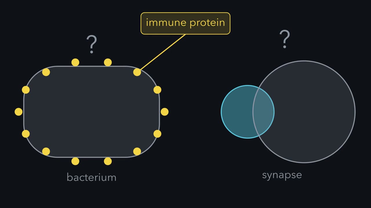
Why can a developing neuron make the first protein of an immune cascade that marks bacteria for destruction?
Continue in the lesson: Microglia and Complement Prune Synapses →
3:04 · Free, with captions and a transcript. The lesson has the full text, the questions and the sources.
More films from this lesson
How this film was made. Narration: an AI narrator voice (Kokoro, af_heart) reading narration written from this lesson's text. Animation: AI-generated in code (Manim) by Claude, a schematic with no drawn molecular structures. Captions come from the narration script. See something wrong? Report it.