Films · Developmental Neuroscience
Where the Line Falls
Read the transcript of “Where the Line Falls”
Film transcript: Where the Line Falls
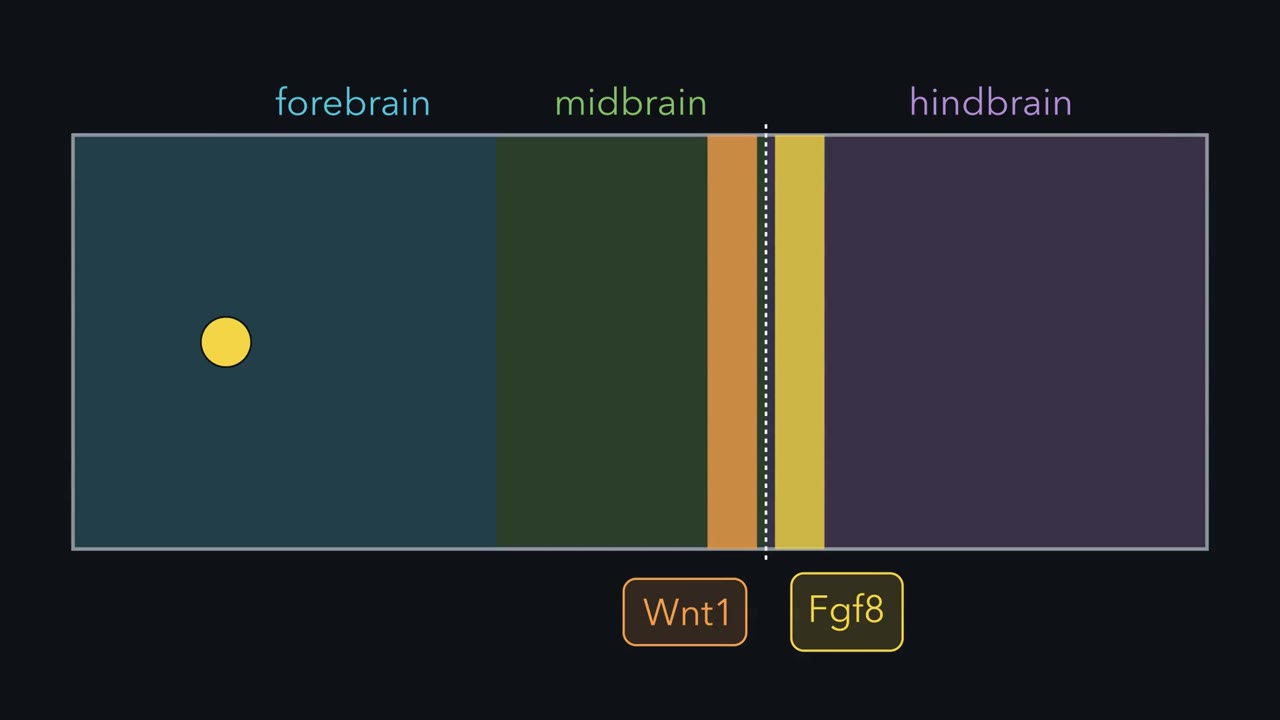
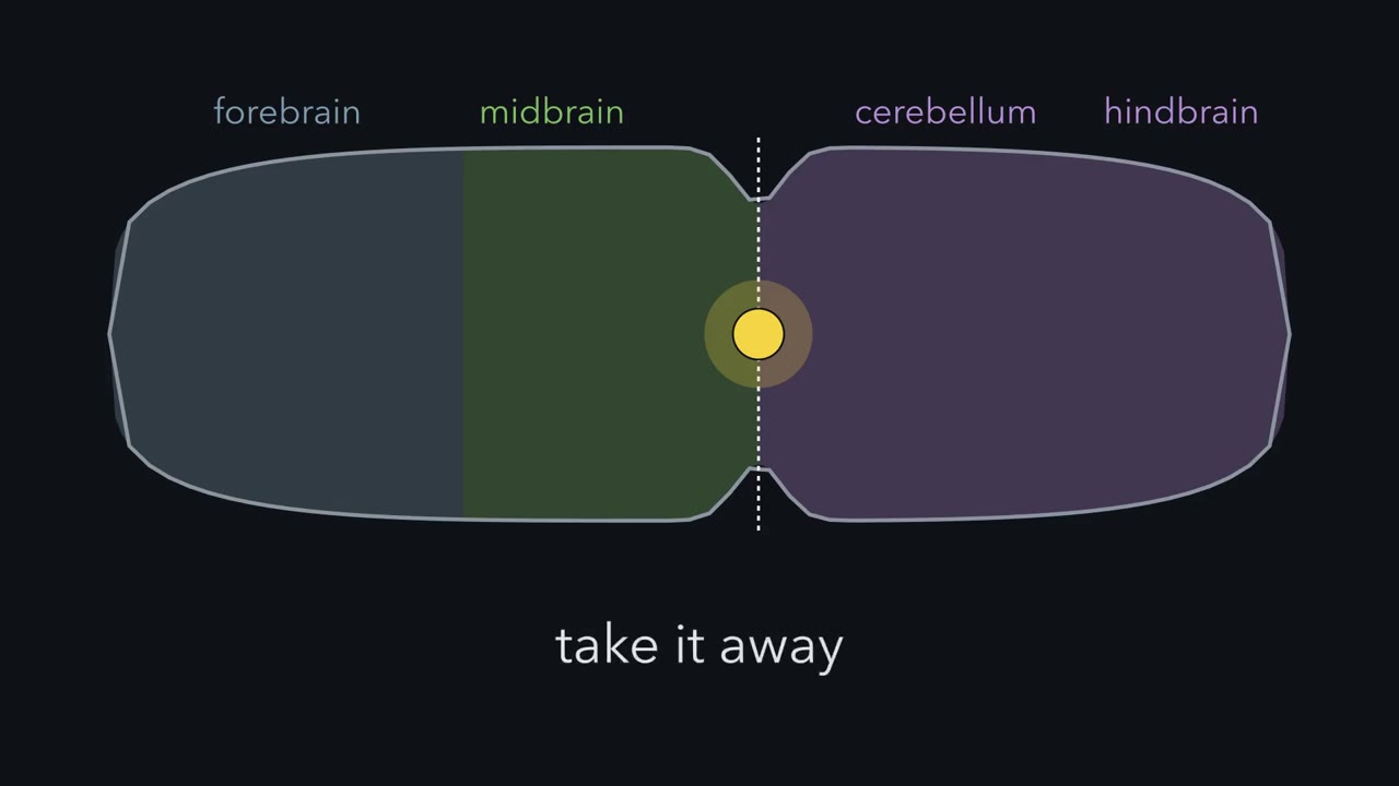
Push a small bead into the back of a chick embryo's forebrain. It is soaked in one protein, FGF8. A second midbrain can grow there, built back to front. Who tells cells what to build? Hox genes give the hindbrain and spinal cord their addresses. The midbrain and forebrain have no Hox code at all. Front at the left: forebrain, then midbrain, then a narrowing called the isthmus. Behind it, the roof of rhombomere 1 gives rise to the cerebellum. A local organizer is a small signaling center inside the tube. It tells nearby cells what to build. The best studied sits at the isthmus. It comes down to one idea. A line becomes a source. Where Otx2 meets Gbx2, the boundary cells become a signaling source.
FGF8 is the chief signal. On the simplest reading, how much signal a cell gets tells it which part to build. The repressor it carries sets which region it can become. In the roof of the tube, that means midbrain in front and cerebellum behind.
Start with the line. Otx2 is on in the forebrain and midbrain. Gbx2 is on in the front of the hindbrain. Each represses the other. A cell with a little more Otx2 makes less Gbx2. Its neighbour does the opposite. So a blurred edge can sharpen, and the line sits where the two meet. How sharp depends on how strong and switch-like the repression is. Predict. In a mouse, force Otx2 on in the front of the hindbrain. Where should the line go? What happens to the cerebellum?
The line moves back, with its band of FGF8. The cerebellum is smaller. Broccoli, Boncinelli and Wurst showed this in 1999. Millet and colleagues did the reverse. Gbx2 made in the back of the midbrain moved the line forward.
This is a move-it test. Change where the players are, and see if the pattern follows. The line follows the repressors. Right at the line, cells make secreted signals: FGF8 just behind it, Wnt1 just in front. This line is more than a fence. Takeaway: two repressors place the line. Next, is that line a source?
How does one protein on a bead make a second midbrain grow, built back to front?
Continue in the lesson: Local Organizers: The Midbrain-Hindbrain Boundary →
2:48 · Free, with captions and a transcript. The lesson has the full text, the questions and the sources.
More films from this lesson
How this film was made. Narration: an AI narrator voice (Kokoro, af_heart) reading narration written from this lesson's text. Animation: AI-generated in code (Manim) by Claude, a schematic with no drawn molecular structures. Captions come from the narration script. See something wrong? Report it.