Films · Developmental Neuroscience
A Tap at the Back, a Drain at the Front
Read the transcript of “A Tap at the Back, a Drain at the Front”
Film transcript: A Tap at the Back, a Drain at the Front
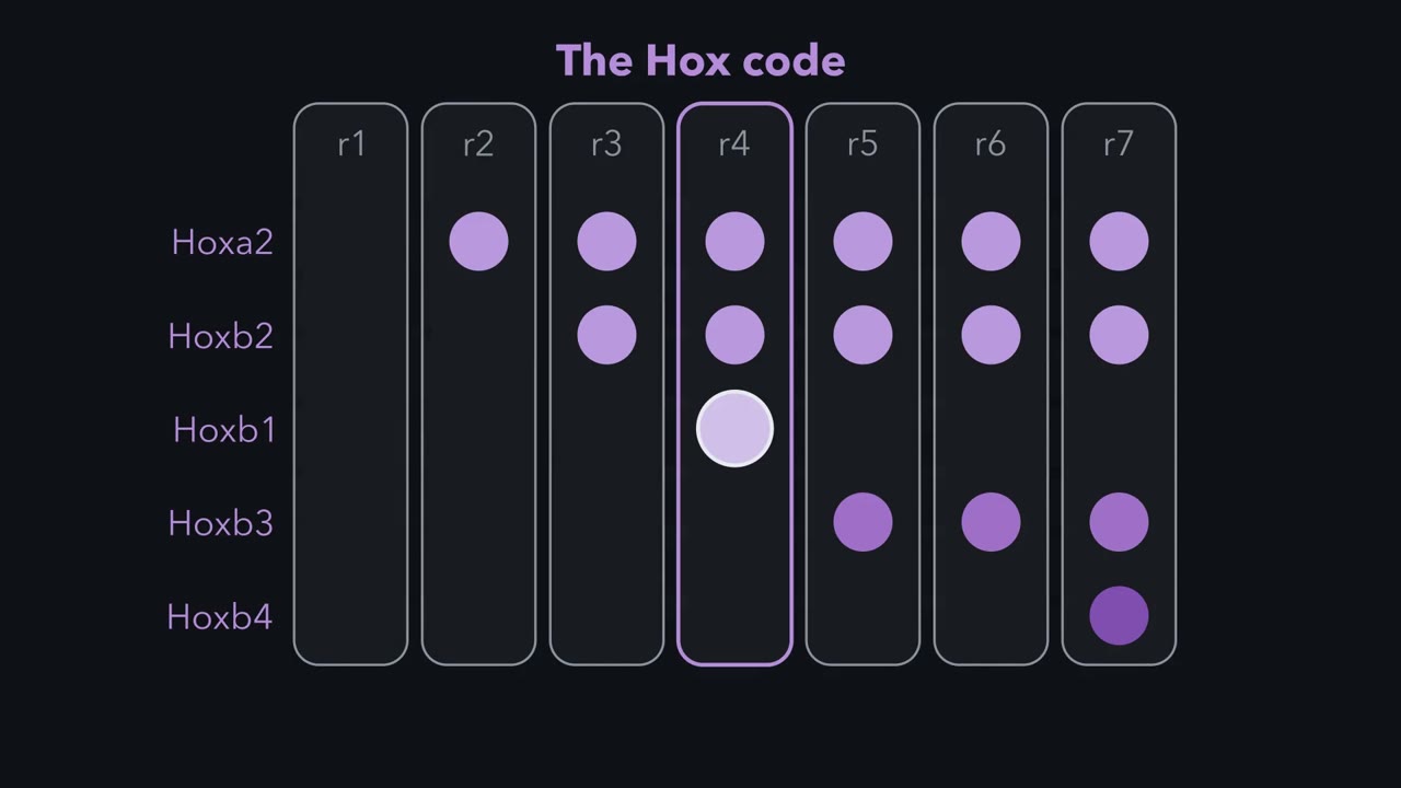
What helps set the front edges of the Hox code? A signal made from vitamin A. Retinoic acid is made from retinol, which is vitamin A, in two steps. Raldh2, the main enzyme for the last step, is made in mesoderm beside the posterior hindbrain and spinal cord. Cyp26 enzymes destroy it. Cyp26a1 is strong toward the front of the neural plate. Picture a bath, with the tap at the back and the drain at the front. Retinoic acid ends up high at the back and low at the front. In the nucleus, it binds the retinoic acid receptor, RAR. RAR pairs with its partner, RXR, on short DNA sequences called response elements. Hoxa1 and Hoxb1 carry these elements. In cell culture, 3′ Hox genes respond to lower doses than 5′ genes.
In a simple threshold model, then, fewer Hox genes come on toward the front, where the signal is lower. Predict. Give mouse embryos extra retinoic acid at an early stage. Which way should the Hox edges move? Forward. In 1992, Marshall, Krumlauf, Lumsden and colleagues found the hindbrain Hox domains changed. Rhombomeres two and three shifted toward the identity of four and five. Too much signal shows a response, not the normal role. Predict. Now block the drain. Mice lacking Cyp26a1 clear less retinoic acid at the front. What changes?
The front shifts: the front of the hindbrain looked partly transformed toward a more posterior character. Remove the tap instead, in mice lacking Raldh2, and the posterior hindbrain is poorly patterned. Changing the signal sets the direction, not the exact edges. In the hindbrain, group 2 genes reach farther forward than group 1. Yet 3′ prime genes need less signal. So other inputs help set those edges. Retinoids are teratogens, agents that cause birth defects. In mouse and chick, extra retinoic acid early shifts the Hox code. It also disturbs the neural crest cells that build much of the face. In humans, this mechanism is inferred from animal work.
More early retinoic acid gives a more posterior code; the code helps set the neurons. Other inputs help draw the final edges.
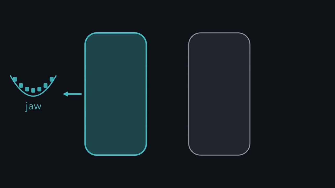
Rhombomeres r2 and r4 start out looking alike. What tells one to build jaw motor neurons and the other facial ones?
Continue in the lesson: Head to Tail: Hox Genes, Retinoic Acid and Rhombomeres →
3:02 · Free, with captions and a transcript. The lesson has the full text, the questions and the sources.
More films from this lesson
How this film was made. Narration: an AI narrator voice (Kokoro, af_heart) reading narration written from this lesson's text. Animation: AI-generated in code (Manim) by Claude, a schematic with no drawn molecular structures. Captions come from the narration script. See something wrong? Report it.