How Microglia Let a Touch Through
Read the transcript of “How Microglia Let a Touch Through”
Film transcript: How Microglia Let a Touch Through
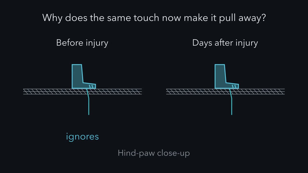
After a nerve injury, a light touch can make a rat pull away. What changed in the spinal cord, and who changed it? Meet a lamina I neuron, in the outermost layer of the dorsal horn. It sends signals toward the brain. In a simple picture, a light touch excites it a little. Inhibitory synapses hold it below threshold. Those synapses open channels that pass chloride. They pull the voltage toward the anion reversal potential. That potential stays low because a transporter, KCC2, carries potassium and chloride out of the cell. In 2003, Coull, De Koninck and colleagues studied rats after nerve injury. Their lamina I neurons had less KCC2. Their anion reversal potential had shifted up.
That does not mean the neuron itself depolarized. Predict. As that potential rises toward threshold, what happens to the inhibitory synapse's grip? Recall the balance-point rule. The synapse can only pull the voltage toward its own reversal potential. So as that potential rises, the pull weakens. Less excitation is needed to fire the cell. In a passive model with made-up values, this touch leaves the steady voltage below threshold in the healthy cell. With the reversal potential at minus −55 mV, it sits exactly at the threshold boundary. At minus −50 mV, it sits above it. Notice what did not happen. Inhibition did not have to turn into excitation.
It only had to weaken. So what changed the neuron? Not a neuron. Tsuda and colleagues, in rats, found P2X4 receptors on reactive microglia. Microglia are the resident immune cells of the cord. They ran two tests. Lowering P2X4 suppressed the allodynia. So the receptor was needed. Microglia stimulated with ATP were transferred into rats never injured. Those rats developed allodynia. So stimulated microglia were enough. The messenger came in 2005. Coull and colleagues showed that stimulated microglia release BDNF. BDNF acts through TrkB receptors. It shifts the anion reversal potential up, the same change seen after injury. Block that signalling, and both the shift and the allodynia reverse.
Three papers, different links, all in rats. The behavioural readout was always a withdrawal threshold. The last link, from weaker inhibition to a touch that fires, is a model. Now the twist. Sorge, Mapplebeck and colleagues gave male and female mice a nerve injury. The hypersensitivity was comparable. In males, a microglial inhibitor reversed the hypersensitivity. In females, under the conditions tested, it did not. Predict. Intact females seemed to rely on adaptive immune cells. What does the inhibitor do in females lacking those cells?
It works. The route can switch. A later study found the same pattern in rats. So it is not a quirk of mice, but it stays conditional. Macrophages in the dorsal root ganglion add another route. In mice, depleting macrophages throughout the body reduced hypersensitivity in both sexes. The authors favoured the ganglion macrophages, but skin dendritic cells were depleted too. So ask of any immune result: which cell, in which sex, in which compartment?
After a nerve injury, a touch the animal used to ignore makes it pull away, and it keeps doing so for weeks. Is persistent pain an alarm that never switched off, or a nervous system that has changed?
Continue in the lesson: When Pain Persists →
4:09 · Free, with captions and a transcript. The lesson has the full text, the questions and the sources.
More films from this lesson
How this film was made. Narration: an AI narrator voice (Kokoro, af_heart) reading narration written from this lesson's text. Animation: AI-generated in code (Manim) by Claude, a schematic with no drawn molecular structures. Captions come from the narration script. See something wrong? Report it.