Developmental Neuroscience · Skim version
Brains in a Dish: Stem Cells and Organoids
Read the full lesson → Skim the whole course →
The guiding idea
Can a person's skin cells, first reset to stem cells and then grown for weeks in a spinning flask, become a model of their early cortex?
The cells bring the program and the embryo brings the surroundings, so an organoid keeps the first and loses most of the second.
From the film: The Program and the Surroundings
- Four transcription factors can reset a skin cell to an induced pluripotent stem cell, which can become any cell type of the body.
- Organoid cells organize themselves into progenitor zones around a cavity with young neurons further out, but the six layers form at best partly and there are no blood vessels.
- The cells bring the program and the embryo brings the surroundings, so an organoid keeps the program and loses most of the surroundings.
- Trust an organoid most for what its cells do on their own, test it hardest where development needs something from outside, and measure every feature.
From the film: Measured Against Fetal Cortex
- With no blood, oxygen must diffuse in from the surface, so in a simple model of a large organoid the core runs out of oxygen and its cells often die.
- Compared cell by cell with fetal cortex, organoids matched the broad classes of cells, such as radial glia, intermediate progenitors and neurons, but finer subtypes were blurred.
- Organoid cells expressed stress genes at higher levels, and those fell after transplant into mouse cortex, which suggests the culture environment contributes.
- The program-and-surroundings rule says where to look first; it is not a verdict, so every feature still has to be measured.
From the film: Putting Back the Surroundings, and Blaming a Mutation
- Fusing a ventral spheroid to a cortical one supplies the missing source of interneurons, which then move into the cortical tissue in jumps.
- In Timothy syndrome assembloids the interneuron migration defect persisted when fused to control cortex, which supports an intrinsic contribution from the interneurons themselves.
- Before blaming a mutation for an organoid phenotype, control for genetic background, cell-line quirks and batch, then put the gene back and take it away.
- Transplanting human organoids into newborn rat cortex supplied a blood supply and responses to whisker deflection.
-
What an organoid is
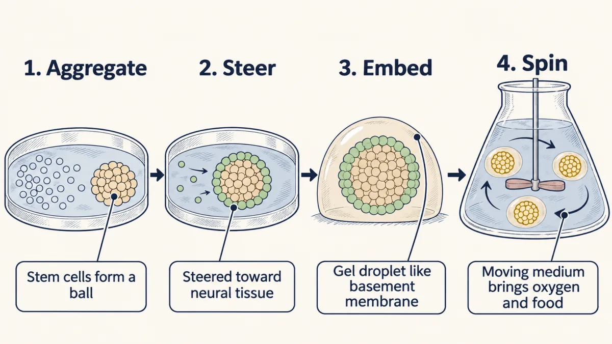
The 2013 cerebral organoid recipe: gather human pluripotent stem cells into balls, steer them toward neural tissue, embed each in a droplet of Matrigel, a gel that resembles basement membrane, and move the droplets to a spinning bioreactor, where moving medium improves the supply of oxygen and nutrients. The cells organize the rest themselves. AI-generated illustration -
Where the cells come from
-
What the cells build on their own
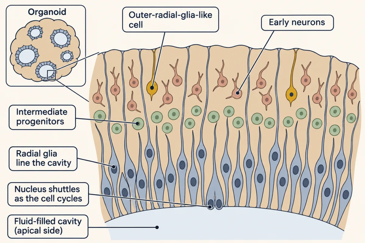
Inside a cerebral organoid, cortex-like zones form around fluid-filled cavities with no one placing the cells: radial glia line the cavity and shuttle their nuclei as they cycle, intermediate progenitors sit beyond them, early neurons lie further out, and outer-radial-glia-like cells, with a basal process and no apical one, are scattered among them. In the 2013 organoids a distinct outer zone like the fetal one was not well formed. AI-generated illustration -
What the dish misses, measured
-
Putting back the surroundings
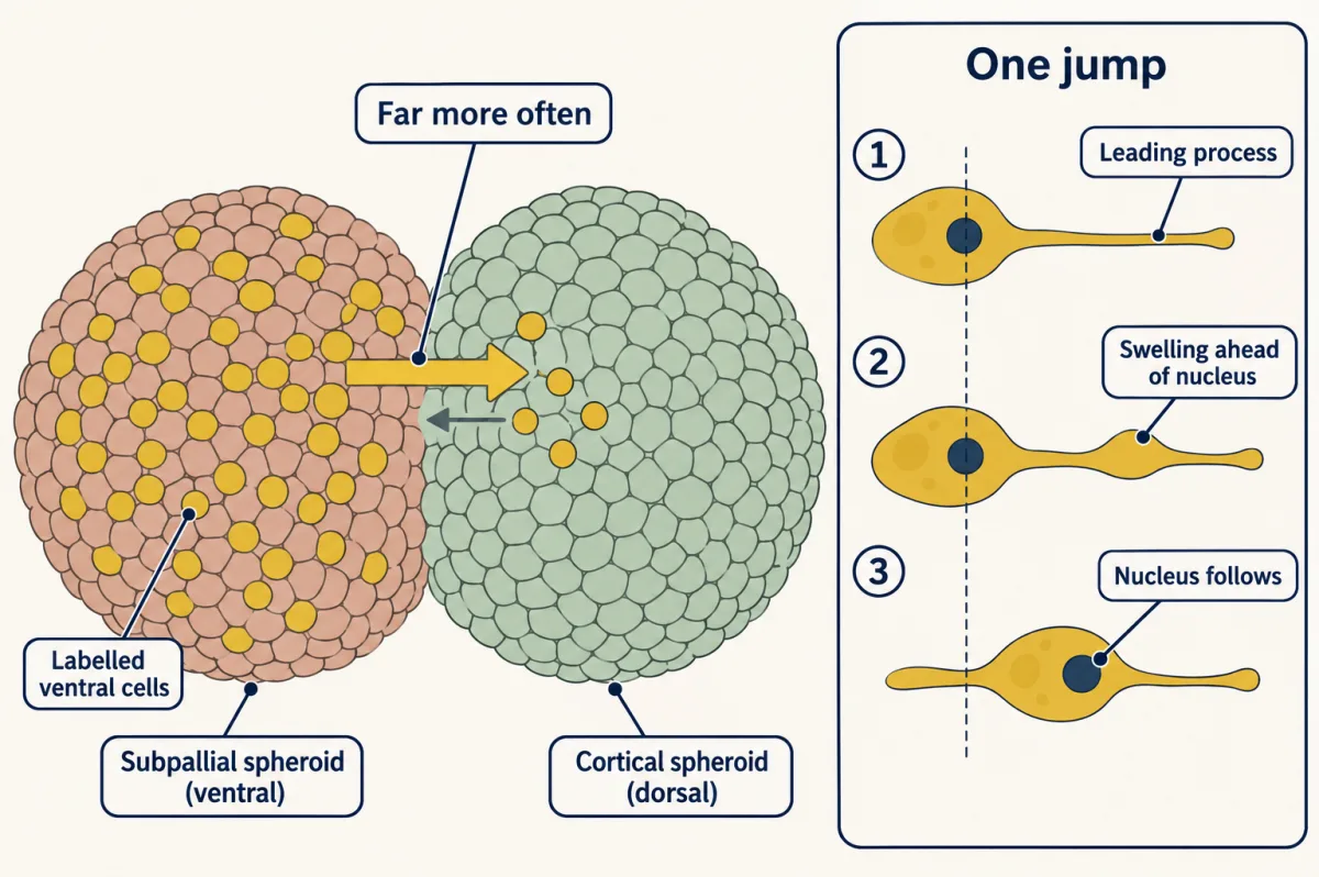
A labelled ventral (subpallial) spheroid fused to a dorsal (cortical) one. Labelled cells cross into the cortical tissue in jumps: leading process, swelling, then nucleus. AI-generated illustration -
Blaming a mutation
-
Putting it together
-
Going deeper
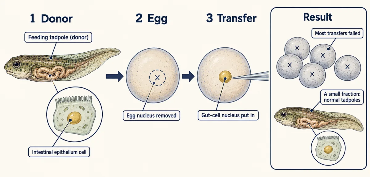
Gurdon put nuclei from the intestinal lining of feeding Xenopus tadpoles into eggs whose own nucleus was removed or destroyed. Most transfers failed, but a small fraction became normal feeding tadpoles. AI-generated illustration
Where this lesson fits
Before thisReview first
- The Ventricular Zone: Radial Glia as Stem CellsThis course · Lesson 13
You need radial glia as cortical stem cells, since organoids form zones of these progenitors around a cavity.
- Tangential Migration: Interneurons and the Long Way RoundThis course · Lesson 19
Interneurons born in ventral forebrain moving into cortex is the process that assembloids are built to study.
- When Building Goes Wrong: Microcephaly, Zika and LissencephalyThis course · Lesson 48
The opening CDK5RAP2 microcephaly result is the disease case that the organoid experiments here test.
Suggested by an AI model from the lessons themselves, then checked. How we use AI