Two Ways to Stop Breathing
Read the transcript of “Two Ways to Stop Breathing”
Film transcript: Two Ways to Stop Breathing
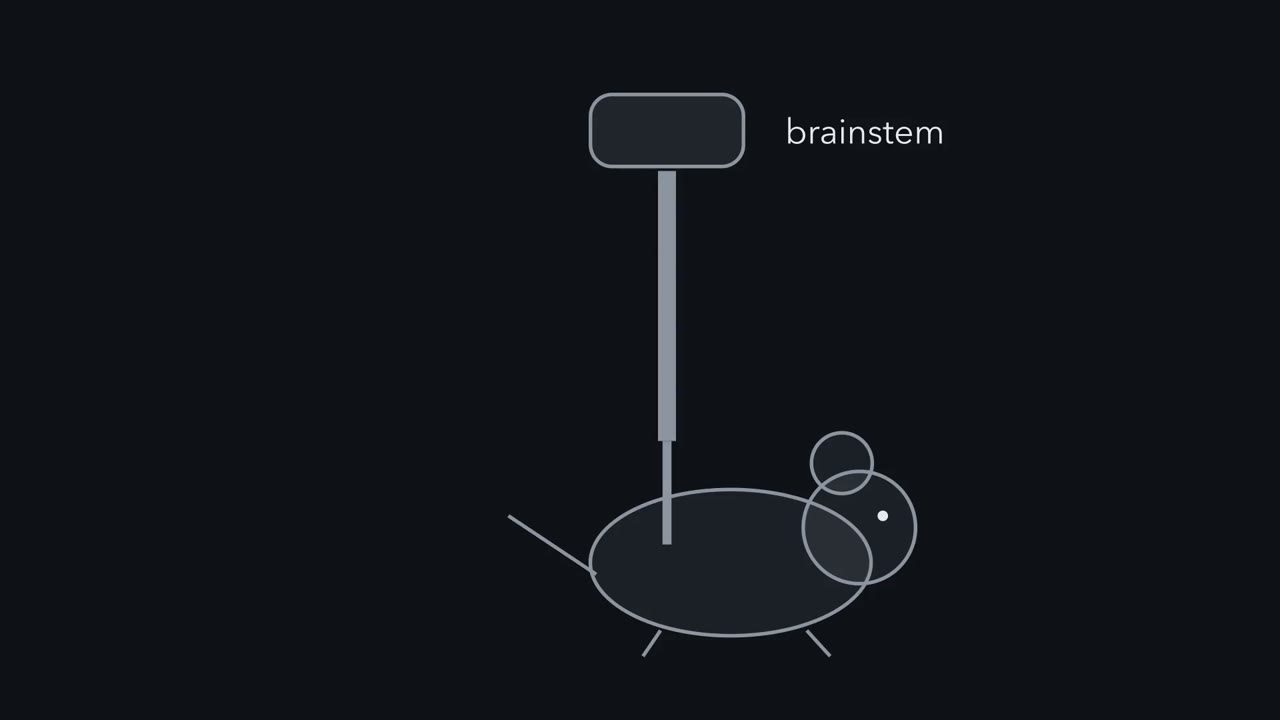
Two findings both end with a mouse not breathing. Are they the same line, used twice? Start with the opposite results. Light on P2ry1 afferents suppressed breathing in anaesthetized mice. Light on Npy2r afferents produced rapid shallow breathing. Opposite outputs, in the same nerve.
This is sufficiency. It says what each population can do when driven, not what drives it in life. Nerve recordings added an independent check. One population was mostly fast, the other mostly slow. A fibres are myelinated and conduct quickly. C fibres are unmyelinated and conduct slowly. Conduction speed was measured apart from the breathing assay. Most of one population conducted fast, most of the other slow. That is a physiological difference, not proof of complete separation.
Now the two routes to one output. First, inflation. When the lungs fill, further inspiration is cut short. This is the Hering-Breuer inflation reflex, described more than a century ago. Cut both vagi and the reflex goes. Second, protection. Water or acid at the larynx triggers protective reflexes that keep the airway clear. Driving P2ry1 afferents with light recruited the whole program, not apnoea alone.
Ablating those cells impaired swallowing to laryngeal water and acid. That is necessity, in that assay. So these cells can recruit airway defence, and one tested piece of it needs them. If they were also the lung's stretch detector, what would inflation imaging show?
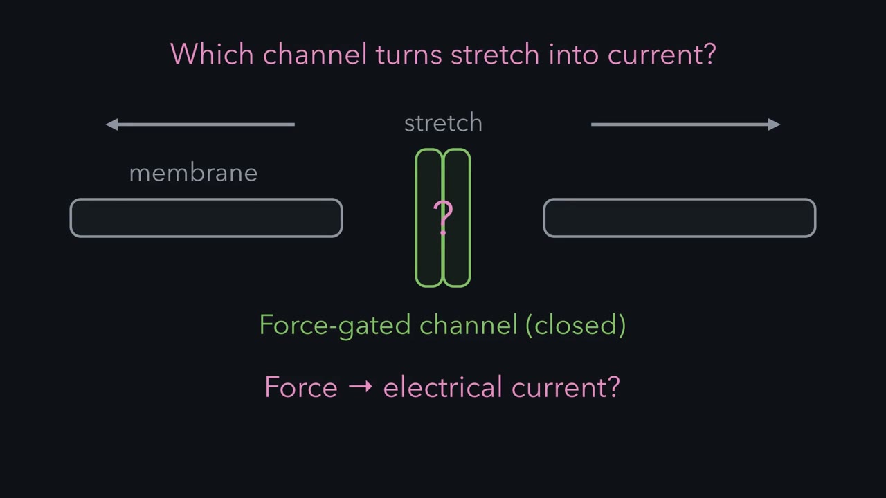
If one population did both jobs, the inflation responders should sit inside the labelled set. They did not. Most stretch-responsive neurons were PIEZO2-positive and P2RY1-negative. That imaging was done with central connections severed, under isoflurane, with the airway inflated in the living animal. So two populations, two challenges, one shared output. The table separates them.
The takeaway is general. An output alone does not identify which pathway produced it. Measure stimulus and output separately.
Light on one set of vagal sensory axons stops an anaesthetized mouse breathing, and light on another set makes it breathe fast and shallow. What does each set report, and how would you know?
Continue in the lesson: Vagal Sensory Neurons: The Body Reports to the Brain →
3:05 · Free, with captions and a transcript. The lesson has the full text, the questions and the sources.
More films from this lesson
How this film was made. Narration: an AI narrator voice (Kokoro, af_heart) reading narration written from this lesson's text. Animation: AI-generated in code (Manim) by Claude, a schematic with no drawn molecular structures. Captions come from the narration script. See something wrong? Report it.