Films · Genetics and Molecular Biology
Reading a PCR as It Grows
Read the transcript of “Reading a PCR as It Grows”
Film transcript: Reading a PCR as It Grows
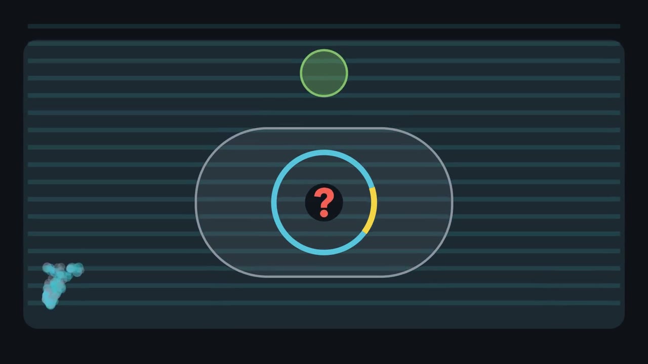
The first film grew DNA inside bacteria. Now two PCR tubes end equally bright. Did they start with the same amount? The answer hides in how the signal grew. Watch a bounded target become a curve that can be read. PCR starts with template DNA, primers, nucleotides, and heat-stable polymerase. A yellow primer binds each grey strand. Their 3' ends point into the target. Polymerase extends from those ends. Primer placement sets the candidate boundaries for amplification. Heating separates template strands. Cooling lets each primer pair with its site. Polymerase extends each primer, building blue DNA along grey template. Repeat the cycle, and earlier products become templates.
Like a photocopier whose copies can be copied, the bounded target branches into a growing family. During ideal early cycles, each completed target can seed another copy. Product can double every cycle, bending the graph upward. The blue curve is computed from starting amount times two raised to cycle number. Real reactions follow this only early. As product accumulates, amplification slows toward a plateau. Different starting amounts can finish at similar levels. Endpoint brightness loses how each reaction arrived there. Real-time PCR preserves that history by measuring fluorescence throughout the run, while early curves remain separated. A green threshold crosses both curves.
One curve reaches it earlier, although both endpoints look alike. Which reaction began with more target? Think about how many rounds each needed to reach the same fluorescence. The earlier crossing came from more starting target. That reaction needed fewer rounds to reach the shared threshold. The crossing is the threshold cycle, written Ct. With matched, near-perfect efficiency, an earlier Ct means more starting template. The plateau cannot support that inference. Real-time quantitative PCR watches DNA accumulate.
Reverse-transcription PCR starts differently. Reverse transcriptase copies RNA into complementary DNA, which PCR can amplify. Combining the steps measures an RNA target. The fluorescence still reports amplified DNA, not RNA directly. A curve is evidence only within a controlled comparison. A tube with no template can expose contamination.
For RNA, a tube without reverse transcriptase can expose genomic DNA. A known positive shows the assay works. Controls turn a glowing curve into an interpretable result. The early rising curve preserves information about starting amount. The endpoint mostly shows that product accumulated. Read the crossing under stated assumptions, then challenge the signal with controls. The useful measurement is when the curve arrives, not how bright it ends.
A white colony survives ampicillin and a PCR tube ends with a bright band. How do you know either contains the DNA you wanted?
Continue in the lesson: Recombinant DNA, Cloning and PCR →
3:48 · Free, with captions and a transcript. The lesson has the full text, the questions and the sources.
More films from this lesson
How this film was made. Narration: an AI narrator voice (Kokoro, af_heart) reading narration written from this lesson's text. Animation: AI-generated in code (Manim) by Claude, a schematic with no drawn molecular structures. Captions come from the narration script. See something wrong? Report it.